By agreement with the publisher, this book is accessible by the search feature, but cannot be browsed.
NCBI Bookshelf. A service of the National Library of Medicine, National Institutes of Health.
Janeway CA Jr, Travers P, Walport M, et al. Immunobiology: The Immune System in Health and Disease. 5th edition. New York: Garland Science; 2001.

Immunobiology: The Immune System in Health and Disease. 5th edition.
Show detailsIn this final part of this chapter we will look at the induced responses of innate immunity. These depend upon the cytokines and chemokines that are produced in response to pathogen recognition. We will therefore start with a brief overview of these proteins, followed by a description of how the macrophage-derived cytokines promote the phagocytic response through recruitment and production of fresh phagocytes and opsonizing molecules, while containing the spread of infection to the bloodstream through the activation of clotting mechanisms. We will also look at the role of the cytokines known as interferons, which are induced by viral infection, and at a class of lymphoid cells, known as natural killer (NK) cells, that are activated by interferons to contribute to innate host defense against viruses and other intracellular pathogens.
The induced innate responses either succeed in clearing the infection or contain it while an adaptive response develops. Adaptive immunity harnesses many of the same effector mechanisms that are used in the innate immune system, but is able to target them with greater precision. Thus antigen-specific T cells activate the microbicidal and cytokine-secreting properties of macrophages harboring pathogens, while antibodies activate complement, act as direct opsonins for phagocytes, and stimulate NK cells to kill infected cells. In addition, the adaptive immune response uses cytokines and chemokines, in a manner similar to that of innate immunity, to induce inflammatory responses that promote the influx of antibodies and effector lymphocytes to sites of infection. The effector mechanisms described here therefore serve as a primer for later chapters on adaptive immunity.
2-19. Activated macrophages secrete a range of cytokines that have a variety of local and distant effects
Cytokines are small proteins (~25 kDa) that are released by various cells in the body, usually in response to an activating stimulus, and induce responses through binding to specific receptors. They can act in an autocrine manner, affecting the behavior of the cell that releases the cytokine, or in a paracrine manner, affecting the behavior of adjacent cells. Some cytokines can act in an endocrine manner, affecting the behavior of distant cells, although this depends on their ability to enter the circulation and on their half-life. Chemokines are a class of cytokines that have chemoattractant properties, inducing cells with the appropriate receptors to migrate toward the source of the chemokine. The cytokines secreted by macrophages in response to pathogens are a structurally diverse group of molecules and include interleukin-1 (IL-1), interleukin-6 (IL-6), interleukin-12 (IL-12), TNF-α, and the chemokine interleukin-8 (IL-8). The name interleukin (IL) followed by a number (for example IL-1, IL-2, and so on) was coined in an attempt to develop a standardized nomenclature for molecules secreted by, and acting on, leukocytes. However, this became confusing when an ever-increasing number of cytokines with diverse origins, structures, and effects were discovered, and although the IL designation is still used, it is hoped that eventually a nomenclature based on cytokine structure will be developed. The cytokines and their receptors are grouped according to their structure in the appendices at the end of this book (cytokines are listed in Appendix III and chemokines in Appendix IV). There are three major structural families: the hematopoietin family, which includes growth hormones as well as many interleukins with roles in both adaptive and innate immunity; the TNF family, which functions in both innate and adaptive immunity and includes some membrane-bound members; and the chemokine family, which we discuss below. Of the macrophage-derived interleukins shown in Fig. 2.31, IL-6 belongs to the large family of hematopoietins, TNF-α is obviously part of the TNF family, while IL-1 and IL-12 are structurally distinct. All have important local and systemic effects that contribute to both innate and adaptive immunity, and these are summarized in Fig. 2.31.

Figure 2.31
Important cytokines secreted by macrophages in response to bacterial products include IL-1, IL-6, IL-8, IL-12, and TNF-α. TNF-α is an inducer of a local inflammatory response that helps to contain infections; it also has systemic effects, (more...)
The recognition of different classes of pathogen may involve signaling through distinct receptors and result in some variation in the cytokines induced. The study of this is still in its infancy, but it is thought to be a way in which appropriate responses can be selectively activated as the released cytokines orchestrate the next phase of host defense. We will see how TNF-α, which is elicited by LPS-bearing pathogens, is particularly important in containing infection by these pathogens, and, how the release of different chemokines can recruit and activate different types of effector cells.
2-20. Chemokines released by phagocytes recruit cells to sites of infection
Among the cytokines released in infected tissue in the earliest phases of infection are members of a family of chemoattractant cytokines known as chemokines. These molecules induce directed chemotaxis in nearby responsive cells and were discovered relatively recently. Because they were first detected in cytokine assays, they were initially named as interleukins: interleukin-8 (IL-8) was the first chemokine to be cloned and characterized, and it remains typical of this family (Fig. 2.32, top). All the chemokines are related in amino acid sequence and their receptors are all integral membrane proteins containing seven membrane-spanning helices. This structure is characteristic of receptors such as rhodopsin (Fig. 2.32, bottom) and the muscarinic acetylcholine receptor, which are coupled to G proteins; the chemokine receptors also signal through coupled G proteins. Chemokines function mainly as chemoattractants for leukocytes, recruiting monocytes, neutrophils, and other effector cells from the blood to sites of infection. They can be released by many different types of cell and serve to guide cells involved in innate immunity and also the lymphocytes in adaptive immunity, as we will learn in Chapters 8–10. Some chemokines also function in lymphocyte development, migration, and angiogenesis (the growth of new blood vessels). The properties of a variety of chemokines are listed in Fig. 2.33; quite why there are so many chemokines is not yet known, and neither is the exact role of each in defense against infection.
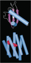
Figure 2.32
Chemokines are a family of proteins of similar structure that bind to chemokine receptors, themselves part of a large family of G protein-coupled receptors. The chemokines are a large family of small proteins represented here by IL-8 (upper molecule). (more...)
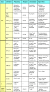
Figure 2.33
Properties of selected chemokines. Chemokines fall mainly into two related but distinct groups: the CC chemokines, which in humans are mostly encoded in one region of chromosome 4, have two adjacent cysteine residues in their amino-terminal region; CXC (more...)
Members of the chemokine family fall mostly into two broad groups—CC chemokines with two adjacent cysteines near the amino terminus, and CXC chemokines, in which the equivalent two cysteine residues are separated by another amino acid. The two groups of chemokines act on different sets of receptors. CC chemokines bind to CC chemokine receptors, of which there are nine so far, designated CCR1–9. CXC chemokines bind to CXC receptors; there are five of these, CXCR1–5. These receptors are expressed on different cell types; in general, CXC chemokines with an Glu-Leu-Arg (ELR) tripeptide motif immediately before the first cysteine promote the migration of neutrophils. IL-8 is an example of this type of chemokine. Other CXC chemokines that lack this motif, such as the B-lymphocyte chemokine (BLC), guide lymphocytes to their proper destination. The CC chemokines promote the migration of monocytes or other cell types. An example is macrophage chemoattractant protein-1 (MCP-1). IL-8 and MCP-1 have similar, although complementary, functions: IL-8 induces neutrophils to leave the bloodstream and migrate into the surrounding tissues; MCP-1, in contrast, acts on monocytes, inducing their migration from the bloodstream to become tissue macrophages. Other CC chemokines such as RANTES may promote the infiltration into tissues of a range of leukocytes including effector T cells (see Section 10-8), with individual chemokines acting on different subsets of cells. The only known C chemokine (with only one cysteine) is called lymphotactin and is thought to attract T-cell precursors to the thymus. A newly discovered molecule called fractalkine is unusual in several ways: it has three amino acid residues between the two cysteines, making it a CX3C chemokine; it is multimodular; and it is tethered to the membrane of the cells that express it, where it serves both as a chemoattractant and as an adhesion protein. We will return to the discussion of chemokines in Chapter 10.
The role of chemokines such as IL-8 and MCP-1 in cell recruitment is twofold. First, they act on the leukocyte as it rolls along endothelial cells at sites of inflammation, converting this rolling into stable binding by triggering a change of conformation in the adhesion molecules known as leukocyte integrins. This allows the leukocyte to cross the blood vessel wall by squeezing between the endothelial cells, as we will see when we describe the process of extravasation. Second, the chemokines direct the migration of the leukocyte along a gradient of the chemokine that increases in concentration toward the site of infection. This is achieved by the binding of the small, soluble chemokines to proteoglycan molecules in the extracellular matrix and on endothelial cell surfaces, thus displaying the chemokines on a solid substrate along which the leukocytes can migrate.
Chemokines can be produced by a wide variety of cell types in response to bacterial products, viruses, and agents that cause physical damage, such as silica or the urate crystals that occur in gout. Thus, infection or physical damage to tissues sets in motion the production of chemokine gradients that can direct phagocytes to sites where they are needed. In addition, peptides that act as chemoattractants for neutrophils are made by bacteria themselves. All bacteria produce proteins with an amino-terminal N-formylated methionine, and, as discovered many years ago, the f-Met-Leu-Phe (fMLP) peptide is a potent chemotactic factor for inflammatory cells, especially neutrophils. The fMLP receptor is also a G protein-coupled receptor like the receptors for chemokines and for the complement fragments C5a, C3a, and C4a. Thus, there is a common mechanism for attracting neutrophils, whether by complement, chemokines, or bacterial peptides. Neutrophils are the first to arrive in large numbers at a site of infection, with monocytes and immature dendritic cells being recruited later. The complement peptide C5a, and the chemokines IL-8 and MCP-1 also activate their respective target cells, so that not only are neutrophils and macrophages brought to potential sites of infection but, in the process, they are armed to deal with any pathogens they may encounter. In particular, neutrophils exposed to IL-8 and the cytokine TNF-α (see Fig. 2.37, and Section 2-23) are activated to produce the respiratory burst that generates oxygen radicals and nitric oxide, and to release their stored lysosomal contents, thus contributing both to host defense and to the tissue destruction and pus formation seen in local sites of infection with pyogenic bacteria.
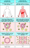
Figure 2.37
The release of TNF-α by macrophages induces local protective effects, but TNF-α can have damaging effects when released systemically. The panels on the left show the causes and consequences of local release of TNF-α, while the (more...)
Chemokines do not act alone in cell recruitment, which also requires the action of vasoactive mediators to bring leukocytes close to the blood vessel endothelium (see Section 2-4) and cytokines such as TNF-α to induce the necessary adhesion molecules on the endothelial cells. We will now turn to the molecules that mediate leukocyte–endothelium adhesion, and then describe the process of leukocyte extravasation step by step, as it is known to occur for neutrophils and monocytes.
2-21. Cell-adhesion molecules control interactions between leukocytes and endothelial cells during an inflammatory response
The recruitment of activated phagocytes to sites of infection is one of the most important functions of innate immunity. Recruitment occurs as part of the inflammatory response and is mediated by cell-adhesion molecules that are induced on the surface of the local blood vessel endothelium. Before we consider the process of inflammatory cell recruitment we will first describe some of the cell-adhesion molecules involved.
A significant barrier to understanding cell-adhesion molecules is their
nomenclature. Most cell-adhesion molecules, especially those on leukocytes,
which are relatively easy to analyze functionally, were named after the effects
of specific monoclonal antibodies against them, and were only later
characterized by gene cloning. Their names therefore bear no relation to their
structure; for instance, the leukocyte functional antigens, LFA-1, LFA-2, and
LFA-3, are actually members of two different protein families.
In Fig. 2.34, the adhesion molecules are
grouped according to their molecular structure, which is shown in schematic
form, alongside their different names, sites of expression, and ligands. Three
families of adhesion molecules are important for leukocyte recruitment. The
selectins are membrane
glycoproteins with a distal lectinlike domain that binds specific carbohydrate
groups. Members of this family are induced on activated endothelium and initiate
endothelial– leukocyte interactions by binding to fucosylated oligosaccharide
ligands on passing leukocytes (see Fig.
8.5). The next step in leukocyte recruitment depends on tighter
adhesion, which is due to intercellular
adhesion molecules (ICAMs) on the endothelium binding to heterodimeric proteins of the
integrin family on leukocytes. We have already encountered two
of the leukocyte integrins that function as complement receptors (CR3 and CR4).
The leukocyte integrins important for extravasation are LFA-1
(αL:β2) and Mac-1 (αM:β2; another name for CR3) and they
bind to both ICAM-1 and ICAM-2 (Fig. 2.35). Strong adhesion between leukocytes and
endothelial cells is promoted by the induction of ICAM-1 on inflamed endothelium
and the activation of a conformational change in LFA-1 and Mac-1 in response to
chemokines. The importance of the leukocyte integrins in inflammatory cell
recruitment is illustrated by the disease leukocyte adhesion deficiency , which stems from a defect in the
β2 chain common to both LFA-1 and Mac-1. People with this disease
suffer from recurrent bacterial infections and impaired healing of wounds.

Figure 2.34
Adhesion molecules in leukocyte interactions. Several structural families of adhesion molecules play a part in leukocyte migration, homing, and cell–cell interactions: the selectins, the integrins, and proteins of the immunoglobulin superfamily. (more...)

Figure 2.35
Phagocyte adhesion to vascular endothelium is mediated by integrins. Vascular endothelium, when it is activated by inflammatory mediators, expresses two adhesion molecules—ICAM-1 and ICAM-2. These are ligands for integrins expressed by phagocytes—α (more...)
The activation of endothelium is driven by interactions with macrophage cytokines, particularly TNF-α, which induces rapid externalization of granules in the endothelial cells called Weibel–Palade bodies. These granules contain preformed P-selectin, which is thus expressed within minutes on the surface of local endothelial cells following production of TNF-α by macrophages. The same effect can be produced directly by exposing cultured human umbilical vein epithelial cells (HUVEC) to LPS, demonstrating that HUVEC can directly sense the presence of infection. Shortly after the appearance of P-selectin on the cell surface, mRNA encoding E-selectin is synthesized, and within 2 hours, the endothelial cells are mainly expressing E-selectin. Both these proteins interact with sulfated-sialyl-Lewisx, which is present on the surface of neutrophils.
Resting endothelium carries low levels of ICAM-2, apparently in all vascular beds. This may be used by circulating monocytes to navigate out of the vessels and into their tissue sites, which happens continuously and essentially ubiquitously. However, upon exposure to TNF-α, local expression of ICAM-1 is strongly induced on the endothelium of small vessels within the infectious focus. This, in turn, binds to LFA-1 on circulating monocytes and polymorpho-nuclear leukocytes, in particular neutrophils, as shown in Fig. 2.35.
Cell-adhesion molecules have many other roles in the body, directing many aspects of tissue and organ development. In this brief description, we have considered only those that participate in the recruitment of inflammatory cells in the hours to days after the establishment of infection.
2-22. Neutrophils make up the first wave of cells that cross the blood vessel wall to enter inflammatory sites
The physical changes that accompany the initiation of the inflammatory response have been described in Section 2-4; here we give a step-by-step account of how the required effector cells are recruited into sites of infection. Under normal conditions, leukocytes are restricted to the center of small blood vessels, where the flow is fastest. In inflammatory sites, where the vessels are dilated, the slower blood flow allows the leukocytes to move out of the center of the blood vessel and interact with the vascular endothelium. Even in the absence of infection, monocytes migrate continuously into the tissues, where they differentiate into macrophages; during an inflammatory response, the induction of adhesion molecules on the endothelial cells, as well as induced changes in the adhesion molecules expressed on leukocytes, recruit large numbers of circulating leukocytes, initially neutrophils and later monocytes, into the site of an infection. The migration of leukocytes out of blood vessels, a process known as extravasation, is thought to occur in four steps. We will describe this process as it is known to occur for monocytes and neutrophils (Fig. 2.36).

Figure 2.36
Neutrophils leave the blood and migrate to sites of infection in a multistep process mediated through adhesive interactions that are regulated by macrophage-derived cytokines and chemokines. The first step (top panel) involves the reversible binding of leukocytes (more...)
The first step involves selectins. P-selectin, which is carried inside endothelial cells in Weibel–Palade bodies, appears on endothelial cell surfaces within a few minutes of exposure to leukotriene B4, the complement fragment C5a, or histamine, which is released from mast cells in response to C5a. The appearance of P-selectin can also be induced by exposure to TNF-α or LPS, and both of these have the additional effect of inducing the synthesis of a second selectin, E-selectin, which appears on the endothelial cell surface a few hours later. These selectins recognize the sulfated-sialyl-Lewisx moiety of certain leukocyte glycoproteins that are exposed on the tips of microvilli. The interaction of P-selectin and E-selectin with these glycoproteins allows monocytes and neutrophils to adhere reversibly to the vessel wall, so that circulating leukocytes can be seen to ‘roll’ along endothelium that has been treated with inflammatory cytokines (see Fig. 2.36, top panel). This adhesive interaction permits the stronger interactions of the next step in leukocyte migration.
This second step depends upon interactions between the leukocyte integrins LFA-1 and Mac-1 with molecules on endothelium such as ICAM-1, which is also induced on endothelial cells by TNF-α (see Fig. 2.36, bottom panel). LFA-1 and Mac-1 normally adhere only weakly, but IL-8 or other chemokines, bound to proteoglycans on the surface of endothelial cells, trigger a conformational change in LFA-1 and Mac-1 on the rolling leukocyte, which greatly increases its adhesive properties. In consequence, the leukocyte attaches firmly to the endothelium and rolling is arrested.
In the third step, the leukocyte extravasates, or crosses the endothelial wall. This step also involves LFA-1 and Mac-1, as well as a further adhesive interaction involving an immunoglobulin-related molecule called PECAM or CD31, which is expressed both on the leukocyte and at the intercellular junctions of endothelial cells. These interactions enable the phagocyte to squeeze between the endothelial cells. It then penetrates the basement membrane (an extracellular matrix structure) with the aid of proteolytic enzymes that break down the proteins of the basement membrane. The movement through the vessel wall is known as diapedesis, and enables phagocytes to enter the subepithelial tissues.
The fourth and final step in extravasation is the migration of leukocytes through the tissues under the influence of chemokines. As discussed in Section 2-20, chemokines such as IL-8 are produced at the site of infection and bind to proteoglycans in the extracellular matrix. They form a matrix-associated concentration gradient along which the leukocyte can migrate to the focus of infection. IL-8 is released by the macrophages that first encounter pathogens and recruits neutrophils, which enter the infected tissue in large numbers in the early part of the induced response. Their influx usually peaks within the first six hours of an inflammatory response, while monocytes can be recruited later, through the action of chemokines such as MCP-1. Once in an inflammatory site, neutrophils are able to eliminate many pathogens by phagocytosis. They act as phagocytic effectors in an innate immune response through receptors for complement and other opsonizing proteins of the innate immune system as well as by recognizing pathogens directly. In addition, as we will see in Chapter 9, they act as phagocytic effectors in humoral adaptive immunity. The importance of neutrophils is dramatically illustrated by diseases or treatments that severely reduce neutrophil numbers. Such patients are said to have neutropenia, and are very susceptible to infection with numerous pathogens. Restoring neutrophil levels in such patients by transfusion of neutrophil-rich blood fractions or by stimulating their production with specific growth factors largely corrects this susceptibility.
2-23. Tumor necrosis factor-α is an important cytokine that triggers local containment of infection, but induces shock when released systemically
Inflammatory mediators also stimulate endothelial cells to express proteins that trigger blood clotting in the local small vessels, occluding them and cutting off blood flow. This can be important in preventing the pathogen from entering the bloodstream and spreading through the blood to organs all over the body. Instead, the fluid that has leaked into the tissue in the early phases of carries the pathogen enclosed in phagocytic cells, especially dendritic cells, via the lymph to the regional lymph nodes, where an adaptive immune response can be initiated. The importance of TNF-α in the containment of local infection is illustrated by experiments in which rabbits are infected locally with a bacterium. Normally, the infection will be contained at the site of the inoculation; if, however, an injection of anti-TNF-α antibody is also given to block the action of TNF-α, the infection spreads via the blood to other organs.
Once an infection spreads to the bloodstream, however, the same mechanisms by which TNF-α so effectively contains local infection instead become catastrophic (Fig. 2.37). The presence of infection in the bloodstream, known as sepsis, is accompanied by the release of TNF-α by macrophages in the liver, spleen, and other sites. The systemic release of TNF-α causes vasodilation and loss of plasma volume owing to increased vascular permeability, leading to shock. In septic shock, disseminated intravascular coagulation (blood clotting) is also triggered by TNF-α, leading to the generation of clots in many small vessels and the massive consumption of clotting proteins, thus causing the patient’s ability to clot blood appropriately to be lost. This condition frequently leads to the failure of vital organs such as the kidneys, liver, heart, and lungs, which are quickly compromised by the failure of normal perfusion; consequently, septic shock has a high mortality rate.
Mice with a mutant TNF-α receptor gene are resistant to septic shock; however, such mice are also unable to control local infection. Although the features of TNF-α that make it so valuable in containing local infection are precisely those that give it a central role in the pathogenesis of septic shock, it is clear from the evolutionary conservation of TNF-α that its benefits in the former area far outweigh the devastating consequences of its systemic release.
2-24. Cytokines released by phagocytes activate the acute-phase response
As well as their important local effects, the cytokines produced by macrophages have long-range effects that contribute to host defense. One of these is the elevation of body temperature, which is mainly caused by TNF-α, IL-1, and IL-6. These are termed endogenous pyrogens because they cause fever and derive from an endogenous source rather than from bacterial components. Fever is generally beneficial to host defense; most pathogens grow better at lower temperatures and adaptive immune responses are more intense at elevated temperatures. Host cells are also protected from the deleterious effects of TNF-α at raised temperatures.
The effects of TNF-α, IL-1, and IL-6 are summarized in Fig. 2.38. One of the most important of these is the initiation of a response known as the acute-phase response (Fig. 2.39). This involves a shift in the proteins secreted by the liver into the blood plasma and results from the action of IL-1, IL-6, and TNF-α on hepatocytes. In the acute-phase response, levels of some plasma proteins go down, while levels of others increase markedly. The proteins whose synthesis is induced by TNF-α, IL-1, and IL-6 are called acute-phase proteins. Several of these are of particular interest because they mimic the action of antibodies, but, unlike antibodies, these proteins have broad specificity for pathogen-associated molecular patterns and depend only on the presence of cytokines for their production.

Figure 2.38
The cytokines TNF-α, IL-1, and IL-6 have a wide spectrum of biological activities that help to coordinate the body’s responses to infection. IL-1, IL-6, and TNF-α activate hepatocytes to synthesize acute-phase proteins, and bone (more...)

Figure 2.39
The acute-phase response produces molecules that bind pathogens but not host cells. Acute-phase proteins are produced by liver cells in response to cytokines released by macrophages in the presence of bacteria. They include serum amyloid protein (SAP) (more...)
One of these proteins, C-reactive protein, is a member of the pentraxin protein family, so called because they are formed from five identical subunits. C-reactive protein is another example of a multipronged pathogen-recognition molecule, and binds to the phosphorylcholine portion of certain bacterial and fungal cell-wall lipopolysaccharides. Phosphorylcholine is also found in mammalian cell membrane phospholipids but in a form that cannot bind to C-reactive protein. When C-reactive protein binds to a bacterium, it is not only able to opsonize it but can also activate the complement cascade by binding to C1q, the first component of the classical pathway of complement activation. The interaction with C1q involves the collagen-like parts of C1q, rather than the globular heads contacted by antibody and pathogen surfaces, but the same cascade of reactions is initiated.
The second acute-phase protein of interest is mannan-binding lectin, which we have already met as a pathogen-binding molecule (see Fig. 2.13) and as a trigger for the complement cascade (see Section 2-7). Mannan-binding lectin is found in normal serum at low levels but is produced in increased amounts during the acute-phase response. It acts as an opsonin for monocytes, which, unlike tissue macrophages, do not express the macrophage mannose receptor. Two other proteins with opsonizing properties that are produced by the liver in increased quantities during the acute-phase response are the pulmonary surfactants A and D. Like mannan-binding lectin and C1q these are members of the collectin family, binding to pathogen surfaces through globular lectin-domains attached to a collagen-like stalk. Pulmonary surfactants A and D are found along with macrophages in the alveolar fluid of the lung and are important in promoting the phagocytosis of respiratory pathogens such as Pneumocystis carinii, one of the main causes of pneumonia in patients with AIDS.
Thus, within a day or two, the acute-phase response provides the host with several proteins with the functional properties of antibodies that bind a broad range of pathogens. However, unlike antibodies, they have no structural diversity and are made in response to any stimulus that triggers the release of TNF-α, IL-1, and IL-6, so their synthesis is not specifically induced and targeted.
A final distant effect of the cytokines produced by phagocytes is to induce a leukocytosis, an increase in circulating neutrophils. The neutrophils come from two sources: the bone marrow, from which mature leukocytes are released in increased numbers; and sites in blood vessels where they are attached loosely to endothelial cells. Thus, the effects of these cytokines contribute to the control of infection while the adaptive immune response is being developed. As shown in Fig. 2.30, TNF-α also has a role in this, as it stimulates the migration of dendritic cells from their sites in peripheral tissues to the lymph node, and their maturation into nonphagocytic but highly co-stimulatory antigen-presenting cells.
2-25. Interferons induced by viral infection make several contributions to host defense
Infection of cells with viruses induces the production of proteins that are known as interferons because they were found to interfere with viral replication in previously uninfected tissue culture cells. They are believed to have a similar role in vivo, blocking the spread of viruses to uninfected cells. These antiviral effector molecules, called interferon-α (IFN-α) and interferon-β (IFN-β), are quite distinct from interferon-γ (IFN-γ). This is not directly induced by viral infection, although it is produced later and does have an important role in the induced response to intracellular pathogens, as we will see below. IFN-α, actually a family of several closely related proteins, and IFN-β, the product of a single gene, are synthesized by many cell types following their infection by diverse viruses. Interferon synthesis is thought to occur in response to the presence of double-stranded RNA, as synthetic double-stranded RNA is a potent inducer of interferon. Double-stranded RNA, which is not found in mammalian cells, forms the genome of some viruses and might be made as part of the infectious cycle of all viruses. Therefore, double-stranded RNA might be the common element in interferon induction.
Interferons make several contributions to defense against viral infection (Fig. 2.40). An obvious and important effect is the induction of a state of resistance to viral replication in all cells. IFN-α and IFN-β are secreted by the infected cell and then bind to a common cell-surface receptor, known as the interferon receptor, on both the infected cell and nearby cells. The interferon receptor, like many other cytokine receptors, is coupled to a Janus-family tyrosine kinase, through which it signals. This signaling pathway, which we will describe in detail in Chapter 6, rapidly induces new gene transcription as the Janus-family kinases directly phosphorylate signal-transducing activators of transcription known as STATs, which translocate to the nucleus where they activate the transcription of several different genes. In this way interferon induces the synthesis of several host cell proteins that contribute to the inhibition of viral replication. One of these is the enzyme oligoadenylate synthetase, which polymerizes ATP into a series of 2′–5′ linked oligomers (nucleotides in nucleic acids are normally linked 3′–5′). These activate an endoribonuclease that then degrades viral RNA. A second protein activated by IFN-α and IFN-β is a serine–threonine kinase called P1 kinase. This enzyme phosphorylates the eukaryotic protein synthesis initiation factor eIF-2, inhibiting translation and thus contributing to the inhibition of viral replication. Another interferon-inducible protein called Mx is known to be required for cellular resistance to influenza virus replication. Mice that lack the gene for Mx are highly susceptible to infection with the influenza virus, whereas genetically normal mice are not.
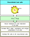
Figure 2.40
Interferons are antiviral proteins produced by cells in response to viral infection. The interferons (IFN)-α and -β have three major functions. First, they induce resistance to viral replication in uninfected cells by activating genes (more...)
Another way interferons protect the host against viruses is by upregulating the cellular immune response to these pathogens. The adaptive immune response to viruses depends on their effective presentation to T cells as peptide fragments complexed with MHC class I molecules at the cell surface, and interferons promote this by inducing increased expression of these molecules. Interferons also activate natural killer (NK) cells to kill virus-infected cells and release cytokines. Although of lymphoid origin, NK cells do not have antigen-specific receptors and are therefore part of the innate immune system. It is not entirely clear what allows them to discriminate between infected and noninfected cells, but they possess both activating and inhibitory receptors. The latter inhibit killing when bound to normal MHC class I molecules and this means that the higher the expression of MHC class I on a cell surface, the more protected it is against destruction by NK cells. Therefore, interferons protect uninfected host cells from NK cells by upregulating class I MHC expression, while activating the NK cells to kill infected cells. Interferons also promote the release of effector cytokines by NK cells, as we will see in the next section.
2-26. Natural killer cells are activated by interferons and macrophage-derived cytokines to serve as an early defense against certain intracellular infections
Natural killer cells (NK cells) develop in the bone marrow from the common lymphoid progenitor cell and circulate in the blood. They are larger than T and B lymphocytes, have distinctive cytoplasmic granules, and are functionally identified by their ability to kill certain lymphoid tumor cell lines in vitro without the need for prior immunization or activation. The mechanism of NK cell killing is the same as that used by the cytotoxic T cells generated in an adaptive immune response; cytotoxic granules are released onto the surface of the bound target cell, and the effector proteins they contain penetrate the cell membrane and induce programmed cell death. However, NK cell killing is triggered by invariant receptors, and their known function in host defense is in the early phases of infection with several intracellular pathogens, particularly herpes viruses, the protozoan parasite Leishmania, and the bacterium Listeria monocytogenes.
NK cells are activated in response to interferons or macrophage-derived cytokines. Although NK cells that can kill sensitive targets can be isolated from uninfected individuals, this activity is increased by between twentyfold and one hundredfold when NK cells are exposed to IFN-α and IFN-β or to the NK cell-activating factor IL-12, which is one of the cytokines produced early in many infections. Activated NK cells serve to contain virus infections while the adaptive immune response generates antigen-specific cytotoxic T cells that can clear the infection (Fig. 2.41). At present the only clue to the physiological function of NK cells in humans comes from a rare patient deficient in NK cells who proved highly susceptible to early phases of herpes virus infection.
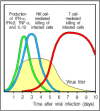
Figure 2.41
Natural killer cells (NK cells) are an early component of the host response to virus infection. Experiments in mice have shown that IFN-α, IFN-β, and the cytokines TNF-α and IL-12 appear first, followed by a wave of NK cells, which together (more...)
IL-12, in synergy with TNF-α, can also elicit the production of large amounts of IFN-γ by NK cells, and this secreted IFN-γ is crucial in controlling some infections before T cells have been activated to produce this cytokine. One example is the response to Listeria monocytogenes. Mice that lack T and B lymphocytes are initially quite resistant to this pathogen; however, antibody-mediated depletion of NK cells or neutralization of TNF-α or IFN-γ or their receptors renders these mice highly susceptible, so that they die a few days after infection before an adaptive immune response can be mounted.
2-27. NK cells possess receptors for self molecules that inhibit their activation against uninfected host cells
If NK cells are to mediate host defense against infection with viruses and other pathogens, they must have some mechanism for distinguishing infected from uninfected cells. Exactly how this is achieved has not yet been worked out, but recognition of ‘altered self’ is thought to be involved. NK cells have two types of surface receptor that control their cytotoxic activity. One type is an ‘activating receptor:’ it triggers killing by the NK cell. Several types of receptor provide this activation signal, including calcium-binding C-type lectins that recognize a wide variety of carbohydrate ligands present on many cells. A second set of receptors inhibit activation, and prevent NK cells from killing normal host cells. These ‘inhibitory receptors’ are specific for MHC class I alleles, which helps to explain why NK cells selectively kill target cells bearing low levels of MHC class I molecules. Altered expression of MHC class I molecules may be a common feature of cells infected by intracellular pathogens, as many of these have developed strategies to interfere with the ability of MHC class I molecules to capture and display peptides to T cells. Thus, one possible mechanism by which NK cells distinguish infected from uninfected cells is by recognizing alterations in MHC class I expression (Fig. 2.42). Another is that they recognize changes in cell-surface glycoproteins induced by viral or bacterial infection.
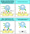
Figure 2.42
Possible mechanisms by which NK cells distinguish infected from uninfected cells. A proposed mechanism of recognition is shown. NK cells can use several different receptors that signal them to kill, including lectinlike activating receptors, or ‘killer (more...)
In mice, inhibitory receptors on NK cells are encoded by a multigene family of C-type lectins called Ly49. Different Ly49 receptors recognize different MHC class I alleles and are differentially expressed on different subsets of NK cells. Some NK cells express Ly49 receptors specific for nonself MHC alleles, but each cell expresses at least one receptor that can recognize an MHC class I allele expressed by the host. In humans, there are inhibitory receptors that recognize distinct HLA-B and HLA-C alleles (these are MHC class I alleles encoded by the B and C loci of the human MHC or Human Leukocyte Antigen gene complex). Although the MHC class I molecules of humans and mice are very similar, these human NK receptors are structurally different from those of the mouse, being members of the immunoglobulin gene superfamily; they are usually called p58 and p70, or killer inhibitory receptors (KIRs). In addition, human NK cells express a heterodimer of two C-type lectins, called CD94 and NKG2. The CD94:NKG2 receptor is also found in mice, and interacts with nonpolymorphic MHC-like molecules, HLA-E in man and Qa-1 in mice, that bind the leader peptides of other MHC class I molecules; thus this receptor may be sensitive to the presence of several different MHC class I alleles. Other inhibitory NK receptors specific for the products of the MHC class I loci are rapidly being defined, and all are members of either the immunoglobulin-like KIR family or the Ly49-like C-type lectins.
Signaling by the inhibitory NK receptors suppresses the killing activity of NK cells. This means that NK cells will not kill healthy genetically identical cells with normal expression of MHC class I molecules, such as the other cells of the body. Virus-infected cells, however, can become susceptible to killing by NK cells by a variety of mechanisms. First, some viruses inhibit all protein synthesis in their host cells, so synthesis of MHC class I proteins would be blocked in infected cells, even while being augmented by interferon in uninfected cells. The reduced level of MHC class I expression in infected cells would make them correspondingly less able to inhibit NK cells through their MHC-specific receptors, and therefore more susceptible to killing. Second, some viruses can selectively prevent the export of MHC class I molecules, which might allow the infected cell to evade recognition by the cytotoxic T cells of the adaptive immune response but would make it susceptible to killing by NK cells. There is also evidence that NK cells can detect the changes in MHC class I molecules that occur when they form complexes with peptides from proteins synthesized as a result of infection, instead of the self peptides from the proteins normally made by the cell. It is not known whether these peptides are recognized directly or whether they alter MHC conformation. Finally, virus infection alters the glycosylation of cellular proteins, perhaps allowing recognition by activating receptors to dominate or removing the normal ligand for the inhibitory receptors. Either of these last two mechanisms could allow infected cells to be detected even when the level of MHC class I expression had not been altered.
Clearly much remains to be learned about this innate mechanism of cytotoxic attack and its physiological relevance. The role of MHC molecules in allowing NK cells to detect intracellular infections is of particular interest as these same molecules govern the response of T cells to intracellular pathogens. It is possible that NK cells, which use a diverse set of nonclonotypic receptors to detect altered MHC, represent the modern remnants of the evolutionary forebears of T cells, which evolved rearranging genes that encode a vast repertoire of antigen-specific receptors geared to recognizing altered MHC.
2-28. Several lymphocyte subpopulations and ‘natural antibodies’ behave like intermediates between adaptive and innate immunity
Receptor gene rearrangements are a defining characteristic of the lymphocytes of the adaptive immune system, and allow the generation of an infinite variety of receptors, each expressed by a different individual T or B cell (see Section 1-10). However, there are several minor lymphocyte subsets that express only a very limited diversity of receptors, encoded by a few common rearrangements. These lymphocytes do not need to undergo clonal expansion before responding effectively to the antigens they recognize, and therefore behave like intermediates between adaptive and innate immunity.
One such group of cells is the intraepithelial subset of γ:δ T cells. The γ:δ T cells are themselves a minor subset of T cells that express receptors that are distinct from the α:β receptors found on the majority of T cells involved in adaptive immunity. They were discovered as a consequence of having immunoglobulin-like receptors encoded by rearranged genes and their function remains obscure. One of their most striking features is their division into two highly distinct sets of cells. One set of γ:δ T cells is found in the lymphoid tissue of all vertebrates and, like B cells and α:β T cells, they display highly diversified receptors. By contrast, intraepithelial γ:δ T cells occur variably in different vertebrates, and commonly display receptors of very limited diversity, particularly in the skin and the female reproductive tract of mice, where the γ:δ T cells are essentially homogeneous in any one site. On the basis of this limited diversity of epithelial γ:δ T-cell receptors and their limited recirculatory behavior, it has been proposed that intraepithelial γ:δ T cells may recognize ligands that are derived from the epithelium in which they reside, but which are expressed only when a cell has become infected. Candidate ligands are heat-shock proteins, MHC class IB molecules, and unorthodox nucleotides and phospholipids, for all of which there is evidence of recognition by γ:δ T cells. Unlike α:β T cells, γ:δ T cells do not generally recognize antigen as peptides presented by MHC molecules; instead they seem to recognize their target antigens directly, and could potentially recognize and respond rapidly to molecules expressed by many different cell types. Recognition of molecules expressed as a consequence of infection, rather than of pathogen-specific antigens themselves, would distinguish γ:δ T cells from other lymphocytes and arguably place them at the intersection between innate and adaptive immunity. However, several recent studies of mice deficient in γ:δ T cells have revealed exaggerated responses to various pathogens and even to self tissues, rather than deficiencies in pathogen control and rejection. This has led to the suggestion that at least some γ:δ T cells have a regulatory role in modulating immune responses, a function that would be consistent with their demonstrated ability to secrete regulatory cytokines when activated. Which aspects of the phenotype of γ:δ-deficient mice are attributable to which subset of γ:δ T cells remains to be clarified.
Another subset of lymphocytes that express a limited diversity of receptors is the B-1 subset of B cells. B cells of this lineage are distinguished by the cell-surface protein CD5 and have properties quite distinct from those of conventional B cells that mediate adaptive humoral immunity. These so-called CD5 B cells, or B-1 cells, are in many ways analogous to epithelial γ:δ T cells: they arise early in ontogeny, they use a distinctive and limited set of gene rearrangements to make their receptors, they are self-renewing in the periphery, and they are the predominant lymphocyte in a distinctive microenvironment, the peritoneal cavity.
B-1 cells seem to make antibody responses mainly to polysaccharide antigens and can produce antibodies of the IgM class without needing T-cell help (Fig. 2.43). Although these responses can be augmented by T cells, they appear within 48 hours of the exposure to antigen, and the T cells involved are therefore not part of an antigen-specific adaptive immune response. The lack of an antigen-specific interaction with helper T cells might explain why immunological memory is not generated: repeated exposures to the same antigen elicit similar or decreased responses with each exposure. Thus, these responses, although generated by lymphocytes with rearranging receptors, resemble innate rather than adaptive immune responses.
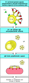
Figure 2.43
CD5 B cells might be important in the response to carbohydrate antigens such as bacterial polysaccharides. These T-cell independent responses occur rapidly, with antibody appearing in 48 hours after infection, presumably because there is a high frequency (more...)
As with γ:δ T cells, the precise role of B-1 cells in host defense is uncertain. Mice that are deficient in B-1 cells are more susceptible to infection with Streptococcus pneumoniae because they fail to produce an antibody against the phospholipid headgroup phosphorylcholine that effectively protects against this organism. A significant fraction of the B-1 cells can make antibodies of this specificity, and because no antigen-specific T-cell help is required, a potent response can be produced early in infection with this pathogen. Whether human B-1 cells have the same role is uncertain.
In terms of evolution, it is interesting to note that γ:δ T cells seem to defend the body surfaces, whereas B-1 cells defend the body cavity. Both cell types are relatively limited in their range of specificities and in the efficiency of their responses. It is possible that these two cell types represent a transitional phase in the evolution of the adaptive immune response, guarding the two main compartments of primitive organisms—the epithelial surfaces and the body cavity. It is not yet clear whether they are still critical to host defense or whether they represent an evolutionary relic. Nevertheless, as each cell type is prominent in certain sites in the body and contributes to certain responses, they must be incorporated into our thinking about host defense.
Finally, there is a collection of antibodies known as ‘natural antibody.’ This ‘natural IgM’ is encoded by rearranged antibody genes that have not undergone somatic mutation. It makes up a considerable amount of the IgM circulating in humans and does not appear to be a result of an antigen-specific adaptive immune response to infection. It has a low affinity for many microbial pathogens, and is very highly cross-reactive, even binding to some self molecules. It is unknown whether this natural antibody has any role in host defense or which type of B cells produce it. Furthermore, it is not known whether it is produced in response to the normal flora of the epithelial surfaces of the body or in response to self. However, it might play a role in host defense by binding to the earliest infecting pathogens and clearing them before they become dangerous.
Summary
Innate immunity can use a variety of induced effector mechanisms to clear an infection or, failing that, to hold it in check until the pathogen can be recognized by the adaptive immune system. These effector mechanisms are all regulated by germline-encoded receptor systems that are able to discriminate between noninfected self and infectious nonself ligands. Thus the phagocytes’ ability to discriminate between self and pathogen controls its release of pro-inflammatory chemokines and cytokines that act together to recruit more phagocytic cells, especially neutrophils, which can also recognize pathogens, to the site of infection. Furthermore, cytokines released by tissue phagocytic cells induce fever, the production of acute-phase response proteins including the pathogen-binding mannan-binding lectin and the C-reactive proteins, and the mobilization of antigen-presenting cells that induce the adaptive immune response. Viral pathogens are recognized by the cells in which they replicate, leading to the production of interferons that serve to inhibit viral replication and to activate NK cells, which in turn can distinguish infected from noninfected cells. As we will see later in this book, cytokines, chemokines, phagocytic cells, and NK cells are all effector mechanisms that also are employed in an adaptive immune response that uses clonotypic receptors to target specific pathogen antigens.
- Activated macrophages secrete a range of cytokines that have a variety of local and distant effects
- Chemokines released by phagocytes recruit cells to sites of infection
- Cell-adhesion molecules control interactions between leukocytes and endothelial cells during an inflammatory response
- Neutrophils make up the first wave of cells that cross the blood vessel wall to enter inflammatory sites
- Tumor necrosis factor-α is an important cytokine that triggers local containment of infection, but induces shock when released systemically
- Cytokines released by phagocytes activate the acute-phase response
- Interferons induced by viral infection make several contributions to host defense
- Natural killer cells are activated by interferons and macrophage-derived cytokines to serve as an early defense against certain intracellular infections
- NK cells possess receptors for self molecules that inhibit their activation against uninfected host cells
- Several lymphocyte subpopulations and ‘natural antibodies’ behave like intermediates between adaptive and innate immunity
- Summary
- Induced innate responses to infection - ImmunobiologyInduced innate responses to infection - Immunobiology
Your browsing activity is empty.
Activity recording is turned off.
See more...