Abstract
Years of use of the antidiabetic drug metformin has long been associated with the risk of vitamin B12 (B12) deficiency in type 2 diabetes (T2D) patients, although the underlying mechanisms are unclear. Accumulating evidence has shown that metformin may exert beneficial effects by altering the metabolism of the gut microbiota, but whether it induces human B12 deficiency via modulation of bacterial activity remains poorly understood. Here, we show that both metformin and the other biguanide drug phenformin markedly elevate the accumulation of B12 in E. coli. By functional and genomic analysis, we demonstrate that both biguanides can significantly increase the expression of B12 transporter genes, and depletions of vital ones, such as tonB, nearly completely abolish the drugs’ effect on bacterial B12 accumulation. Via high-throughput screens in E. coli and C. elegans, we reveal that the TetR-type transcription factor RcdA is required for biguanide-mediated promotion of B12 accumulation and the expressions of B12 transporter genes in bacteria. Together, our study unveils that the antidiabetic drug metformin helps bacteria gather B12 from the environment by increasing the expressions of B12 transporter genes in an RcdA-dependent manner, which may theoretically reduce the B12 supply to T2D patients taking the drug over time.
Subject terms: Cellular microbiology, Molecular medicine
The antidiabetic drug, metformin, helps E. coli gather vitamin B12 from the environment by increasing the expressions of transporter genes in a RcdA-dependent manner.
Introduction
Currently, metformin is still the first-line pharmaceutical option for treating type 2 diabetes mellitus (T2D) worldwide1,2. However, despite many beneficial effects proposed for the use of metformin, it is found that long-term use of the drug commonly results in B12 deficiency in T2D patients, with a prevalence up to 41%3–5. Individuals with B12 deficiency usually exhibit neuropathy with symptoms, such as ataxia, diminished cognition, hematologic abnormalities of microcytosis and megaloblastic anemia6. Specifically, metformin-induced low B12 status has been linked with a reduction in cognitive function and an increase in the risk of depression, which are generally found to be irreversible upon the occurrence of demyelination and nerve degeneration7–9.
B12 is indispensable for cellular growth and homeostasis due to its essential roles in DNA synthesis and metabolism of amino acids as well as fatty acids10. Humans and the majority of small-intestinal microbes are completely dependent on a dietary source of B12, as they lack B12 biosynthesis genes11,12. Dietary B12 is initially released in the stomach, where it binds to intrinsic factor to form the complex followed by binding with the cubilin receptor on the distal ileum, eventually being sent to the liver for storage13. The amount of B12 stored in the liver is usually sufficient to meet physiological needs, which has made difficult to experimentally induce B12-deficient models for mechanistic studies14,15. Likely, at least in part, due to the lack of sound B12-deficient models, the mechanisms underlying metformin-induced B12 deficiency remain largely unknown.
Numerous lines of evidence suggest that metformin exerts its beneficial effects, including antihyperglycemic and antiaging effects, by altering the gut microbiome16–20. Intriguingly, it has been reported that biguanide-associated B12 deficiency can be reversed by the administration of antibiotics21, indicating a potential role of bacteria in biguanide-induced B12 malabsorption in humans. It is noted that the uptake of metformin and B12 in the host occurs through the distal ileum, where a considerable amount of B12 auxotroph gut microbes exist and provides a platform for potential interactions among the drug, B12 and microbes. A recent study also indicated that bacterial B12 transporters may help the microbiota compete with the host for B12 by dissociating the host intrinsic factor-B12 complex22. Moreover, the depletion of B12 by the gut microbiota has been proposed as a primary cause of B12 malabsorption in certain human disorders, such as blind loop syndrome23,24. However, whether metformin influences the host B12 level through gut microbes remains unknown.
Unlike the symbiotic relationship between humans and gut microbes, C. elegans directly eats bacteria as a source of food, making it a suitable tool to detect B12 levels in bacteria25,26. In the present study, through functional analysis of B12 activity in worms and direct measurement of bacterial B12 levels, we demonstrated that biguanides significantly increased the amount of B12 in multiple E. coli strains. Interestingly, such B12 accumulation induced by biguanides was found to be fully dependent on the activity of functional B12 transporters. Mechanistically, we found that both biguanides could increase the expressions of certain vital B12 transporter genes in E. coli, especially tonB, suggesting a possible mechanism by which biguanides promote B12 accumulation in bacteria. Through a genome-wide screen with the E. coli Keio collection library for genes responsible for biguanide action using B12-sensing worm models25–27, among which the tonB mutant bacterial clone was included as a positive control, we revealed that the biofilm formation process and transmembrane transport were the top enriched pathways. Knockout of genes from those enriched pathways significantly impaired the effect of phenformin in elevating bacterial B12 levels. By conducting another round of narrow-down filtering, we further identified the transcription factor RcdA, a TetR-type transcription factor28,29, as the element responsible for biguanide-increased B12 accumulation in E. coli. Moreover, we demonstrated that RcdA was required for the biguanide-mediated increase in the expressions of corresponding B12 transporter genes, indicating that RcdA acts upstream of B12 transporters in response to metformin exposure. Altogether, our study not only provides insights into the mechanisms underlying the effect of biguanide drugs in helping bacteria capture B12 from the environment but also lays a foundation for understanding the role of the gut microbiota in B12 deficiency induced by long-term use of metformin in T2D patients.
Results
Metformin and phenformin promote bacterial B12 accumulation
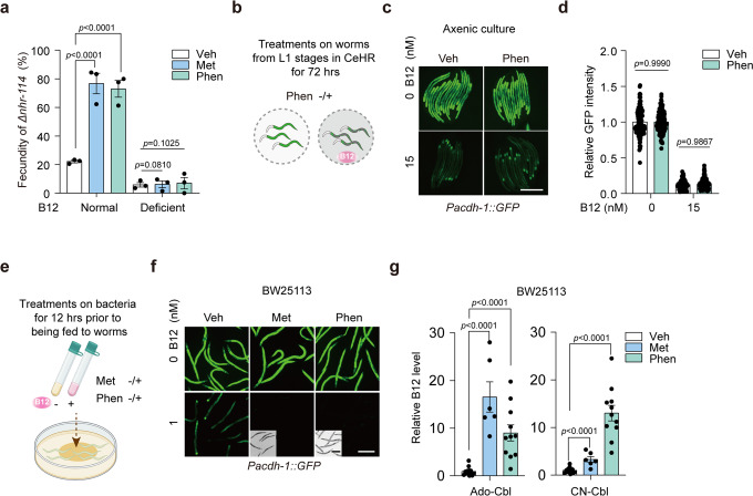
To investigate whether biguanides can change the level of B12 in bacteria, we employed the nhr-114 loss-of-function (hereafter abbreviated as ∆nhr-114) worm model, which is a newly proposed B12-deficient model developed by us and others25,27. Phenotypically, these animals exhibit fertility in a B12 dose-dependent manner. Interestingly, we found that the infertility-rescuing effect of biguanides was completely dependent on the presence of B12, which disappeared when drugs were supplied under B12-deficient conditions (Fig. 1a). To determine whether biguanides elevate the levels of B12 in worms through bacteria, we used the well-established B12 sensor Pacdh-1::GFP worms, whose GFP intensity can indicate the level of B12 present in the diet26,30,31. We administered phenformin to Pacdh-1::GFP worms in an axenic culture system (Fig. 1b–d) or fed the sensor worms with drug-pretreated E. coli BW25113 (Fig. 1e, f), another widely-used E. coli strain for mechanistic studies32–34. Surprisingly, we found that biguanides increased the levels of B12 in the B12 sensor worms only when the drugs were administered in the presence of bacteria, suggesting that biguanides promoted B12 accumulation in bacteria rather than worms directly. To possibly mimic the physiological condition in patients where bacteria are exposed to a long-term treatment with low-dose biguanides, we explored whether a lower dose of phenformin could induce bacterial B12 accumulation in a relatively longer treatment compared to the conditions mainly applied in our study. Indeed, we found that B12 levels in bacteria were increased by 2 mM phenformin with time (Supplementary Fig. 1), suggesting that such bacterial B12 absorption increase may occur in T2D patients during the long-term and low-dose therapy with metformin.
Fig. 1. The antidiabetic biguanides promote bacterial B12 accumulation.
a Fecundity of Δnhr-114 animals on normal NGM plates or B12-deficient NGM plates treated with 50 mM metformin/5 mM phenformin. Veh, vehicle. Met, metformin. Phen, phenformin. N = 3 independent experiments containing at least 30 worms per group. b Scheme of treatments on worms in axenic culture treated with B12 and/or phenformin. c Representative images of Pacdh-1::GFP worms supplemented with B12 and/or 4 mM phenformin in axenic culture. d Quantification of relative GFP intensity of Pacdh-1::GFP worms in (c). N = 3 independent experiments containing at least 30 worms per condition. e Scheme of treatments on BW25113 to feed Pacdh-1::GFP worms. f Representative images of Pacdh-1::GFP worms fed with BW25113 pretreated with B12 and/or 200 mM metformin/4 mM phenformin. N = 3 independent experiments containing at least 30 worms per condition. Scale bar: 250 μm (fluorescence images) and 500 μm (bright field images) for (c) and (f). g LC-MS/MS measurement of B12 levels in BW25113 treated with 200 mM metformin/4 mM phenformin. Ado-Cbl, adenosylcobalamin; CN-Cbl, cyanocobalamin. N = 2 independent experiments for the metformin groups and N =3 independent experiments with at least 3 single colonies per test. The statistical significance values were determined by ordinary one-way ANOVA for (a) and (d) and unpaired t test for (g). Error bars denoted the S.E.M.
To examine the exact extent to which the B12 levels were changed by biguanides in bacteria, we employed liquid chromatography with tandem mass spectrometry (LC-MS/MS) to measure B12 levels in bacteria with or without drug treatment. The results showed that both drugs significantly increased the accumulation of B12 in the adenosylcobalamin (Ado-Cbl) and cyanocobalamin (CN-Cbl) forms in bacteria (Fig. 1g). Theoretically, the CN-Cbl form is the exogenously supplemented version of B12, while the Ado-Cbl form should be converted from CN-Cbl inside of live bacteria35,36. Interestingly, we found that phenformin at 4 mM induced a much higher accumulation of CN-Cbl but a lower level of Ado-Cbl compared to those outcomes induced by metformin at 200 mM (Fig. 1g), which may indicate a more potent activity of phenformin in blocking the bacterial conversion of Ado-Cbl from CN-Cbl than metformin as reported in other applications37,38. Together, these results demonstrated that biguanides promote bacterial B12 accumulation from the environment.
The B12 transport system is required for phenformin-induced bacterial B12 accumulation
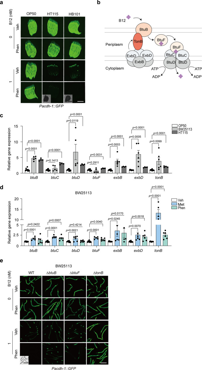
We administered phenformin with concomitant B12 or not to the E. coli strains that are typically used in C. elegans studies, including OP50, HT115, and HB101, to explore whether the drug could elevate the accumulation of B12 in different bacterial types. The treated bacteria were then examined by feeding to the B12 sensor worms and using LC-MS/MS measurement. Indeed, we found that phenformin strongly induced B12 accumulation in the K12 x B hybrid strain HB101 and the K12 strain HT115 (Fig. 2a and Supplementary Fig. 2a). However, it appeared that the B strain OP50 showed a weaker capacity to accumulate B12 from the environment than other strains either treated by phenformin or not, consistently indicated by the GFP intensities of B12 sensor animals and LC-MS/MS measurement on B12 levels (Fig. 2a and Supplementary Fig. 2a).
Fig. 2. The B12 transport machinery plays essential roles in phenformin-mediated bacterial B12 accumulation.
a Representative images of Pacdh-1::GFP worms fed bacterial strains OP50, HT115, and HB101 pretreated with B12 and/or 4 mM phenformin. N = 3 independent experiments containing at least 30 worms per group. b Diagram of the B12 transport machinery in E. coli. c–d Relative mRNA levels of genes involved in B12 transport in OP50, BW25113, and HT115 bacteria (c) and BW25113 with 200 mM metformin/4 mM phenformin treatment or not (d). For (c, d), N = 2 independent experiments containing 4–6 replicates. The statistical significance values were determined by multiple t tests. Error bars denoted the S.E.M. e Representative images of Pacdh-1::GFP worms fed WT and B12 transporter mutant strains pretreated with B12 and/or 4 mM phenformin. WT, BW25113. N = 3 independent experiments containing at least 30 worms per group. Scale bar: 250 μm (fluorescence images) and 500 μm (bright field images) for (a) and (e).
It has been documented that E. coli requires an active transport system to absorb B12 from the environment for growth and survival39,40. B12 is normally transported across the outer membrane by BtuB into the periplasm, which is dependent on the energy transduced from the TonB complex, composed of TonB, ExbB, and ExbD. In the periplasm, B12 is captured and delivered by BtuF to the ATP-binding-cassette transporter BtuCD and ultimately released into the cytoplasm (Fig. 2b). Thus, we speculated that the weak B12 accumulation capacity in the OP50 strain may be attributed to the low activity or expression of genes in the control of B12 transport. Intriguingly, we found that the OP50 strain showed significantly lower expression of all the tested genes for B12 transport than BW25113 or HT115 (Fig. 2c), which explained its poor performance in absorbing B12 compared to that of other strains and the weakened effect of phenformin (Fig. 2a and Supplementary Fig. 2a). Considering the strong association between the activity of B12 transporters and the intrinsic capacity of bacterial B12 absorption, we wondered whether the B12 transport gene expression was altered by phenformin treatment. Indeed, we found that both metformin and phenformin induced the expressions of most of the B12 transporter genes, among which tonB was the most significant one (Fig. 2d). Furthermore, we confirmed that the deletion of tonB, btuB and btuF could markedly abolish the effect of phenformin on the induction of B12 accumulation in BW25113 (Fig. 2e), although not all the tested genes were from the B12 transport system (Supplementary Fig. 2b). Collectively, our results suggested that the B12 transport system, likely via its gatekeeping function, is essential for phenformin-induced B12 levels in E. coli.
High-throughput screens for genes mediating the action of biguanides in bacteria
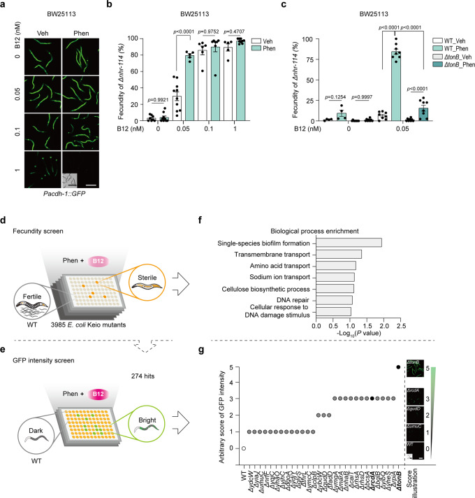
To identify the genetic machinery responsible for bacterial B12 accumulation by biguanides, we set up genetic screens using Δnhr-114 animals whose reproduction could be fully restored by B1225,27 and the B12 sensor worm strain Pacdh-1::GFP. To better design the screens, we compared the sensitivities of these two worm models upon supplementation with various doses of B12 and found that the Δnhr-114 readout showed higher sensitivity to the low dose of B12 than the GFP sensor strain, especially when 0.05 nM B12 was co-administered with phenformin to the bacteria (Fig. 3a, b). To evaluate the effect of bacterial mutants on worm reproduction, we included the tonB mutant strain as a positive control, in which basal B12 absorption and phenformin-mediated B12 induction were both found to be impaired (Fig. 2e). We confirmed that the effect of phenformin in inducing B12 accumulation in bacteria was significantly reduced in ΔtonB according to the low fecundity of Δnhr-114 (Fig. 3c and Supplementary Fig. 3a). Therefore, we carried out the primary unbiased screen with 3,985 bacterial mutants starting with Δnhr-114 fecundity readout (Fig. 3d and Supplementary Fig. 3b), followed by a secondary screen employing the B12 sensor animals Pacdh-1::GFP to narrow down the hits (Fig. 3e). From the primary screen, we identified 274 candidate genes, the knockout of which significantly reduced the effect of phenformin on bacterial B12 accumulation (Supplementary Data 1). Gene Ontology analysis revealed that biofilm formation and transmembrane transport were the most significantly enriched biological processes (Fig. 3f and Supplementary Data 1). From the secondary screen, 28 candidate genes were screened out, among which ΔtonB obtained the highest score (Fig. 3g and Supplementary Fig. 3c).
Fig. 3. Bacterial genetic screens to identify the effectors of phenformin with worm B12 sensors.
a Representative images of Pacdh-1::GFP worms fed BW25113 pretreated with various doses of B12 and/or 4 mM phenformin. N = 3 independent experiments containing at least 30 worms per group. b The fecundity of Δnhr-114 worms fed BW25113 pretreated with various doses of B12 and/or 2 mM phenformin. N = 5 independent experiments containing 1–2 replicates per experiment. c The fecundity of Δnhr-114 worms fed WT and ΔtonB bacteria pretreated with 0.05 nM B12 and/or 2 mM phenformin. N = 4 independent experiments containing 1–2 replicates each time. The statistical significance values were determined by two-way ANOVA for (b, c). Error bars denoted the S.E.M. d, e Schemes of genetic screens for genes responsible for phenformin-mediated bacterial B12 accumulation. Fecundity screen (d). The cartoons of sterile and fertile Δnhr-114 worms were adapted from our previous work25. The whole library was screened using Δnhr-114 worms treated with 2 mM phenformin and 0.05 nM B12. N = 4 independent experiments containing at least 30 worms per well and each round. GFP intensity screen (e). A total of 274 hits from the fecundity screen were screened using Pacdh-1::GFP worms treated with 4 mM phenformin and 1 nM B12. N = 3 independent experiments containing at least 30 worms per well and each round. f Biological process enrichment analysis of 274 hits from the fecundity screen. g The fluorescence scores of Pacdh-1::GFP worms on WT and 28 hits from the GFP intensity screen. Scale bar: 250 μm (fluorescence images) and 500 μm (bright field images) for (a) and (g).
Biguanides induce both bacterial growth inhibition and B12 accumulation by modulating RcdA
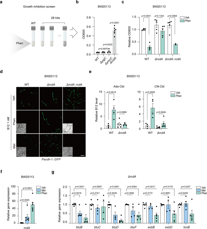
To identify the target genes of biguanides that play the most consistent roles in the drug actions from the 28 hits, we included another trait of biguanides, bacterial growth inhibition (Fig. 4a), the phenotype of which has been considered to be closely associated with other effects of biguanides41. We first validated the growth inhibition effects of phenformin in four different E. coli strains (Supplementary Fig. 4), and then explored if such effects were bacteriostatic or bactericidal. The results showed that 5 mM phenformin treatment retarded the growth of E. coli, and slightly reduced the number of living cells, indicating that phenformin inhibited bacterial growth but did not kill them (Supplementary Fig. 5a). Moreover, 5 mM phenformin indeed induced dual effects on bacterial growth inhibition according to their cell density and integrity indicated by OD600 measurement and dsDNA binding dye SYBR Green I staining, respectively (Supplementary Fig. 5b, c). Collectively, we concluded that the growth inhibition effect of phenformin was mainly bacteriostatic.
Fig. 4. Biguanides induce bacterial growth inhibition and B12 accumulation through RcdA.
a Scheme of the growth inhibition screen. The growth statuses of 28 hits from the secondary screen after treatment with 5 mM phenformin were evaluated by optical density. b The outcomes of the growth inhibition screen. OD600 value measurement of WT, ΔyqjC, ΔymcC, and ΔrcdA. N = 2 independent experiments containing 6 replicates. c Growth of WT, ΔrcdA, and ΔrcdA::rcdA cells treated with 4 mM phenformin. N = 2 independent experiments containing 2–5 replicates. d Representative images of Pacdh-1::GFP worms fed WT, ΔrcdA, and ΔrcdA::rcdA pretreated with 4 mM phenformin/200 mM metformin or not. N = 3 independent experiments containing at least 30 worms per group. Scale bar: 250 μm (fluorescence images) and 500 μm (bright field images). e LC-MS/MS measurement of B12 levels in WT and ΔrcdA treated with 4 mM phenformin or untreated. N = 3 independent experiments containing 4–5 replicates. f Relative mRNA levels of rcdA with 200 mM metformin or 4 mM phenformin treatment. g Relative mRNA levels of genes involved in B12 transport in ΔrcdA with 200 mM metformin or 4 mM phenformin treatment. For (f, g), N = 2 independent experiments containing 4–6 replicates. The statistical significance values were determined by ordinary one-way ANOVA for (b) and (e) and multiple t tests for (c) and (f, g). Error bars denoted the S.E.M.
We reasoned that deletion of the drug target genes would confer the mutant strains resistance to high concentrations of phenformin. Interestingly, only 3 out of the 28 candidates could grow up under phenformin treatment, among which ∆rcdA exhibited the most significant resistance to the drug (Fig. 4b). Moreover, RcdA is a transcription factor controlling the expression of many important genes involved in biofilm formation28, which were candidates out of initial screens (Fig. 3f), suggesting that RcdA might be a key factor determining the effect of biguanides in inducing bacterial B12 accumulation. To test whether RcdA mediated the effects of phenformin by modulating drug accumulation, we measured the drug levels in ∆rcdA using LC-MS/MS. The results showed that the deletion of rcdA did not alter the amount of phenformin accumulated in bacteria compared to that in WT (Supplementary Fig. 6), indicating that RcdA mediated the activity of the drug instead of its level in bacteria.
To confirm the dual role of RcdA in mediating the effects of phenformin on both bacterial growth inhibition and B12 accumulation, we re-expressed the RcdA protein in the rcdA mutant strain. The complementary expression of RcdA nearly completely restored the bacterial sensitivity and B12-accumulating property in response to biguanide treatments (Fig. 4c, d). By using LC-MS/MS, we more directly measured the B12 levels in WT and ΔrcdA with or without drug treatment. Consistent with the results indicated by the B12 sensor animals (Fig. 4d), the deletion of rcdA significantly reduced the effect of phenformin in promoting B12 accumulation in both the Ado-Cbl and CN-Cbl forms compared to WT (Fig. 4e). We then examined the gene expression of rcdA and found it significantly increased upon treatments with both biguanides (Fig. 4f). Collectively, these results indicated that biguanides promote bacterial B12 accumulation by increasing the expression of rcdA.
To explore whether the deletion of rcdA abolished the effects of biguanides by modulating B12 transporters, we tested the expressions of B12 transporter genes in ∆rcdA with or without biguanide treatments. The results showed that neither metformin nor phenformin could upregulate the expression of those genes in ∆rcdA as they did in WT (Figs. 2d, 4g). Surprisingly, both biguanides instead even reduced the expression of btuB or btuD in ∆rcdA through a yet unknown mechanism (Fig. 4g). It implied that there might be a parallel pathway regulating the expressions of B12 transporter genes under biguanide treatments independent of RcdA. Together, our results suggested that biguanides elevate bacterial B12 uptake from the environment by promoting the expressions of B12 transporter genes, which presumably leads to B12 shortage in the environment, the host, over time (Supplementary Fig. 7).
Discussion
Evident B12 deficiency has been routinely found to be associated with a range of symptoms, including diarrhea, anemia, and irreversible neuropathy42,43. More frequently than in other settings, B12 deficiency in patients taking metformin remains unrecognized, possibly due to its gradual development. Accordingly, there is no definitive guideline for the treatment of B12 deficiency in patients taking metformin thus far44, despite B12 supplementation generally prescribed for relieving the possible resulting consequences. A better understanding of the mechanisms underlying metformin-induced B12 deficiency would contribute to the development of guidelines for the diagnosis and treatment of this specific type of B12 deficiency.
Using the model organism E. coli, our study demonstrated that metformin increases bacterial B12 accumulation from the environment. Through a combination of different screens with worms and E. coli, we mechanistically unveiled the molecular basis by which biguanide drugs exert their effects in helping bacterial B12 accumulation, i.e., by transcriptionally elevating the expressions of B12 transporter genes in an RcdA-dependent manner. Altogether, the results of our study provide a perspective for further investigating the action of metformin in inducing B12 deficiency in humans and a foundation for developing more targeted interventions against such side effects.
Metformin has been consistently suggested to be beneficial in promoting longevity and treating metabolic disorders in worms and humans through modulation of gut bacterial metabolism16,18,45. A recent study reported that metformin exerts immediate effects on changes of the composition and function of gut microbes in T2D patients. They found that the drug treatment is accompanied with a significant enrichment of genes responsible for bacterial environmental responses, such as ATP-binding-cassette transporters16. Other groups also found that metformin treatment can alter bacterial membrane function and activities in varied bacteria species46,47. There are a variety of bacteria colonizing the human distal ileum48, the majority of which lack enzymes for de novo B12 biosynthesis and rely on the uptake of exogenous B1249. These bacteria have been reported with high capacities in obtaining B12 from the colonized host by modulating B12 transporters50,51. Here, we demonstrated that both biguanides could elevate B12 accumulation in multiple E. coli strains by increasing the expressions of B12 transporters in an RcdA-dependent manner. Whether biguanides can elicit such an effect in other species of gut microbiota warrants future studies.
The transcription factor RcdA is known with activities in mediating functions of the biofilm master regulator CsgD28 and other players involved in stress responses29. Notably, the biofilm formation pathway was enriched in our initial screens, although depletions of those genes seemed no effects on modulating phenformin’s growth inhibition activity in bacteria (Fig. 4b), indicating a potential role of biofilm formation in bacterial B12 gathering. RcdA is recently found in complex with multiple ligands likely to sense or respond to environmental cues52. Furthermore, our study proposed a previously unappreciated role of RcdA in mediating biguanide-induced bacterial B12 accumulation. The transport of B12 in E. coli is thought mainly relied on the Btu system and the energy-transducing TonB complex53. There are a number of ways reported to regulate the B12 transporter genes, including metal-dependent regulators, σ/anti-σ factor systems, small RNAs on TonB complex and the riboswitch regulation on btuB54,55. Whether and how biguanides modulate the expression of B12 transporter genes through these pathways, in a RcdA-dependent or -independent mode, warrant further investigations.
It is worth mentioning that this study presented an acute treatment model of metformin action using relatively high doses of the drug compared to its long-term and low-dose therapy in T2D patients. Our results showed that both metformin and phenformin can markedly promote bacterial B12 accumulation from the culture environment, but whether they elevate B12 accumulation in the physiological context is worthy of future research. Alternatively, a biguanide-induced B12-deficient mouse model, which is outside of our current expertise, can be applied in future studies to validate and advance our findings toward potential clinical applications.
Methods
Bacterial strains
The E. coli strains OP50, HT115 and HB101 were obtained from the Caenorhabditis Genetics Center (CGC). The Keio Knockout Collection (Cat# OEC4988) and the parent strain BW25113 (Cat#OEC5042) were purchased from Dharmacon. E. coli strains were grown at 37 °C in normal LB or B12-deficient medium (tryptone replaced by neutralized soy-peptone, named soy medium thereafter). E. coli deletion mutants were grown at 37 °C in normal LB or soy medium with 50 μg/mL kanamycin56.
C. elegans strains
Worms were maintained as described at 20 °C57. The strains VC1760 (nhr-114(gk849)) and VL749 (wwIs24 [Pacdh-1::GFP + unc-119(+)]) were purchased from the CGC. All experiments were performed with synchronized hermaphroditic animals.
Chemicals
CN-Cbl (Sigma Aldrich, Cat# V900445) was dissolved in ddH2O as a stock solution at 15 mM. Metformin and phenformin (Sigma Aldrich, Cat# D150959 and Cat# PHR157) were prepared in ddH2O as stock solutions at 1 M and 0.2 M, respectively. Varied doses of biguanides were applied in this study according to previous studies18,38 and the level of tolerance of tested organisms to the drugs. In brief, 50 mM metformin was used in the on-plate treatment for ∆nhr-114 worms, and 200 mM was used in all bacterial treatments. The same volumes of ddH2O were used as vehicle (Veh). Phenformin at 4 mM was used in all bacterial treatments for feeding Pacdh-1::GFP worms, LC-MS/MS measurement, growth measurement and qPCR. Phenformin was used at an optimized concentration, 2 mM, in the Keio Collection screen for feeding Δnhr-114 worms to ensure sufficient bacterial growth. The highest dose of phenformin (5 mM) was used in on-plate treatments or growth inhibition assays to screen for the strongest phenformin-resistant strains. All reagents were filtered with 0.22 μm membranes prior to use and stored at −20 °C.
Fecundity assessment of Δnhr-114 worms
E. coli OP50 was cultured in normal LB or soy medium and seeded on NGM or B12-deficient NGM (peptone was replaced by neutralized soya peptone, OXIOD, Cat# LP0044T) agar plates. The plates were treated with metformin and phenformin at the indicated concentrations and ddH2O as Veh for 4 h before dropping the L1s of Δnhr-114 worms. Alternatively, E. coli strains were cultured in soy medium with the indicated treatments at 37 °C for 12 h. The pretreated bacterial liquid was concentrated and seeded on B12-deficient NGM agar plates before dropping the L1s. The fecundity of the worms was quantified at the day 2 stage58. Animals were counted as fertile (with eggs in the uterus) or sterile (no eggs). The fecundity ratio was calculated as
| 1 |
Imaging of Pacdh-1::GFP worms
E. coli strains were cultured in soy medium with the indicated treatments at 37 °C for 12, 24 or 48 h. The pretreated bacterial liquid was concentrated and seeded on B12-deficient NGM agar plates. Pacdh-1::GFP worms in the L1 stage were dropped on the plates and cultured until the L4 stage. They were collected, anesthetized in M9 containing levamisole (1 mg/mL) and mounted on glass slides for imaging. All images were taken with a Leica DM500 microscope with fixed exposure parameters.
Axenic medium culture
The B12-deficient C. elegans Habituation and Reproduction (CeHR) medium was prepared using a recipe (Supplementary Data 2) modified from others59. Synchronized Pacdh-1::GFP worms in the L1 stage were cultured in CeHR medium supplemented with B12 at indicated concentrations with continuous shaking at 70 rpm on a shaker at 20 °C. Synchronization was conducted according to the hypochlorite method to remove bacteria.
LC-MS/MS measurement of B12 and phenformin
The levels of B12 and phenformin in E. coli were determined by LC-MS/MS. E. coli was cultured in 10 mL soy medium supplemented with 1 nM CN-Cbl and 4 mM phenformin or 200 mM metformin treatment for 12 h. Bacteria were collected by centrifugation and then washed with sterile M9 buffer 3 times. The bacteria were resuspended in 450 μL ddH2O, and the samples were stored at −80 °C overnight. Samples were thawed and frozen with liquid nitrogen 3 times and lysed with the sonication device (Sonics VCX150 Ultrasonicator) using the program 10 s on 10 s off, for 5 mins, on ice. After sonication, the samples were centrifuged for 20 min to pellet the debris. The supernatant was transferred to new tubes, and the protein was precipitated with acetonitrile:methanol:supernatant (67.5:22.5:10 vol/vol/vol). The samples were then centrifuged at the maximum speed for 30 min at 4 °C. The supernatant was collected, lyophilized for 5 h, and resuspended in 50 µL ddH2O for LC-MS/MS detection. The concentrations of B12 and phenformin were normalized to the total cell numbers.
LC-MS/MS analysis was conducted using an AB SCIEX QTRAP 6500+ mass spectrometer with an Exion LC system. Chromatographic separation was achieved on an ACQUITY UPLC BEH C18 column (100 mm × 2.1 mm, 1.7 μm) at 40 °C. The mobile phase consisted of water containing 0.1% formic acid, 20 mM ammonium acetate (A) and methanol (B) at a flow rate of 0.4 mL/min. The gradient of mobile phase B was 3% for 1 min, ramping up from 3% to 60% in 4 min, holding at 60% for 1 min, ramping up from 60% to 95% in 0.1 min, holding at 95% for 1.9 min, ramping down from 95% to 3% in 0.1 min, and holding at 3% for 1.9 min. The injected volume for each sample was 3 μL. The mass spectrometer was operated in positive ion mode using the following settings: curtain gas 40 psi, ion spray voltage 4.5 kV, source temperature 550 °C, ion source gas 1 60 psi, and ion source gas 2 55 psi. Compounds were measured by multiple reaction monitoring (MRM) with optimized cone voltage and collision energy.
RNA extraction and quantitative RT–PCR
The expressions of bacterial genes were evaluated by RT-qPCR. For gene expression evaluations, bacteria were inoculated in 3 mL regular LB medium at the ratio of 1% from overnight starters for 12 h. Specifically, for drug treated groups, bacteria were cultured in 3 mL soy medium supplemented with 1 nM CN-Cbl and 4 mM phenformin or 200 mM metformin for 12 h. Total RNA was extracted with RNAzol (GeneCopoeia, Cat# QP020) with bacterial pellets. gDNA was removed with DNase before reverse transcription with 5x Hiscript® III QRT SuperMix (Vazyme, Cat# R323-01). Quantitative PCR was conducted with SYBR Green PCR reagent (Vazyme, Cat# Q711-02) on a quantitative PCR system (Jena Qtower 3 G). For comparison in different bacterial strains, the expression levels of genes of interest were normalized to that of idnT, while those from the drug-treated groups were normalized to that of rrsA whose expression was not affected by biguanides. The sequences of the primers used are listed in Supplementary Data 3.
E. coli deletion mutant screen with Δnhr-114 worms
Keio E. coli deletion collection clones were grown overnight in 500 μL of soy medium containing 0.05 nM B12, 2 mM phenformin, and 50 μg/mL kanamycin in 96-well deep-well plates. The overnight bacterial culture was concentrated to 25 μL (30 μL for those wells on the margin) and seeded onto B12-deficient 96-well plates. Approximately 30 synchronized L1 of Δnhr-114 animals were placed onto each well of the plates, and worms were allowed to develop for 96–120 h at 20 °C before observation. The fecundity phenotype in each well was scored from 0 (sterile) to 5 (fertile), as noted in Supplementary Data 1.
Genotyping of bacterial deletion strains
Single gene deletion bacterial strains were streaked onto LB plates containing 50 μg/mL kanamycin. PCR was carried out on individual colonies using genomic and kanamycin-cassette-specific primers. Genomic primers were designed for individual strains starting 100–1000 bases upstream of the start codon of the gene and 100–1000 bases downstream of the stop codon60. PCR products were analyzed for the correct sizes separated by agarose gel electrophoresis. The sequences of the primers used in this study were listed in Supplementary Data 3.
Bacterial colony forming assay
Bacterial viability was evaluated by comparing the colony forming units (C.F.U.) of bacterial cultures prior to and post phenformin treatment. The assay was performed with an initial bacterial culture containing 1 x 10⁶ CFU bacteria/mL, which was diluted from overnight grown bacteria. The untreated sample was directly from the initial culture, and the phenformin-treated one obtained from the bacteria treated by the drug at 5 mM for another 12 h in 37 °C, with continuously shaking at 220 rpm. Both samples were diluted and streaked on LB agar plates followed by incubation at 37 °C for 12 h. The number of colonies formed were counted and the relative C.F.U. was calculated by
| 2 |
Extracellular DNA (eDNA) staining by SYBR Green I
Single colonies of BW25113 were inoculated and cultured in LB medium for 12 h as starters of the staining assay. These overnight cultures were aliquoted and treated with or without 5 mM phenformin for 6 or 12 h at 37 °C. Samples from each time point were collected for OD600 measurement and stained with SYBR Green I (MCE, Cat# HY-K1004, 10,000x). Images of bacteria post SYBR Green I staining were acquired with Olympus FV3000-BX63 with 63x immersion objective and analyzed by ImageJ software. More than 3 images per slide were taken for fluorescence quantification.
Bacterial growth inhibition assay
Bacteria were cultured in 200 μL LB supplemented with 5 mM phenformin in 96-well flat-bottom plates at 950 rpm for 12 h. The optical density (OD) of the overnight bacterial culture was obtained by using a plate reader (Thermo Varioskan LUX Microplate reader) at a wavelength of 600 nm. The experiment was performed in duplicate and repeated two times.
Construction of the complementary strain
To construct the plasmids for ΔrcdA complementation, the rcdA fragment was amplified and then ligated into the pUC57::amp plasmid (Addgene#196258). The generated recombinant vector (pUC57::amp::rcdA) was subsequently transformed into the competent ΔrcdA mutant. The complementation strain loaded with pUC57::amp::rcdA was selected on an LB plate containing ampicillin. Finally, the selected complementation strain was validated by PCR and designated ΔrcdA::rcdA.
Statistics and reproducibility
GraphPad Prism 8.0 (GraphPad Software, Inc.) was used for statistical analyses in this study. All results were obtained from at least three biologically replicates. For experiments using C. elegans, at least 30 worms were analyzed for each condition. For differences between two groups, unpaired Student’s t test was used. For differences among multiple groups, one-way analysis of variance (ANOVA) followed by Tukey’s test was used. Two-way ANOVA was used to analyze differences between multiple groups with two variations. p < 0.05 was considered to be statistically significant. Data are presented as the mean ± S.E.M. The statistical significance of differences was inserted in each figure.
Reporting summary
Further information on research design is available in the Nature Portfolio Reporting Summary linked to this article.
Supplementary information
Description of Additional Supplementary Files
Acknowledgements
The authors thank Jinheng Pan, Jia Chen, Shan Feng, Zhenzhen Yu, and Yalin Wang for facility support and the C. elegans Genetics Center (CGC) for providing strains. This work was supported by the Westlake Education Foundation and the National Natural Science Foundation of China (32071151).
Author contributions
L.W., L.Y., and Y.W. together conceived and designed the study. L.Y. and Y.W. both conducted experiments and data analysis and wrote a draft of the work; S.Q. contributed to experiments with Δnhr-114 animals and related discussions. S.Z. contributed to the axenic culture experiments. L.W. supervised the project and wrote the manuscript with input from all authors.
Peer review
Peer review information
Communications Biology thanks Man Kit Cheung, Min Cao and Janis Klovins for their contribution to the peer review of this work. Primary Handling Editors: Sridhar Mani and George Inglis. Peer reviewer reports are available.
Data availability
The authors declare that all data of this study have been provided within the article and Supplementary data set. The source data for all presented figures were provided in Supplementary Data 4. Further information, resources, and reagents are available from the corresponding author upon reasonable request.
Competing interests
The authors declare no competing interests.
Footnotes
Publisher’s note Springer Nature remains neutral with regard to jurisdictional claims in published maps and institutional affiliations.
These authors contributed equally: Luxia Yao, Yihan Wang.
Supplementary information
The online version contains supplementary material available at 10.1038/s42003-023-04475-0.
References
- 1.Foretz M, Guigas B, Bertrand L, Pollak M, Viollet B. Metformin: from mechanisms of action to therapies. Cell Metab. 2014;20:953–966. doi: 10.1016/j.cmet.2014.09.018. [DOI] [PubMed] [Google Scholar]
- 2.Soukas AA, Hao H, Wu L. Metformin as anti-aging therapy: is it for everyone? Trends Endocrinol. Metab. 2019;30:745–755. doi: 10.1016/j.tem.2019.07.015. [DOI] [PMC free article] [PubMed] [Google Scholar]
- 3.Beulens JWJ, Hart HE, Kuijs R, Kooijman-Buiting AMJ, Rutten GEHM. Influence of duration and dose of metformin on cobalamin deficiency in type 2 diabetes patients using metformin. Acta Diabetol. 2015;52:47–53. doi: 10.1007/s00592-014-0597-8. [DOI] [PubMed] [Google Scholar]
- 4.Tomkin GH, H D W J Vitamin-B12 status of patients on long-term metformin therapy. Br. Med. J. 1971;5763:685–687. doi: 10.1136/bmj.2.5763.685. [DOI] [PMC free article] [PubMed] [Google Scholar]
- 5.Chapman LE, Darling AL, Brown JE. Association between metformin and vitamin B12 deficiency in patients with type 2 diabetes: A systematic review and meta-analysis. Diabetes Metab. 2016;42:316–327. doi: 10.1016/j.diabet.2016.03.008. [DOI] [PubMed] [Google Scholar]
- 6.Ahmed MA, Muntingh GL, Paul R. Perspectives on peripheral neuropathy as a consequence of metformin-induced vitamin B12 deficiency in T2DM. Int. J. Endocrinol. 2017;2017:1–6. doi: 10.1155/2017/2452853. [DOI] [PMC free article] [PubMed] [Google Scholar]
- 7.Biemans E, et al. Cobalamin status and its relation with depression, cognition and neuropathy in patients with type 2 diabetes mellitus using metformin. Acta Diabetol. 2015;52:383. doi: 10.1007/s00592-014-0661-4. [DOI] [PubMed] [Google Scholar]
- 8.Goodarzi, Mark O. Increased risk of cognitive impairment in patients with diabetes is associated with metformin. Diabetes Care. 2014;36:2981–2987. doi: 10.2337/dc13-0229. [DOI] [PMC free article] [PubMed] [Google Scholar]
- 9.Stabler SP. Clinical practice. Vitamin B12 deficiency. N. Engl. J. Med. 2013;368:149–160. doi: 10.1056/NEJMcp1113996. [DOI] [PubMed] [Google Scholar]
- 10.Green R, et al. Vitamin B12 deficiency. Nat. Rev. Dis. Prim. 2017;3:17040. doi: 10.1038/nrdp.2017.40. [DOI] [PubMed] [Google Scholar]
- 11.Fang H, Kang J, Zhang D. Microbial production of vitamin B12: a review and future perspectives. Microb. Cell Fact. 2017;16:15. doi: 10.1186/s12934-017-0631-y. [DOI] [PMC free article] [PubMed] [Google Scholar]
- 12.LeBlanc JG, et al. Bacteria as vitamin suppliers to their host: a gut microbiota perspective. Curr. Opin. Biotech. 2013;24:160–168. doi: 10.1016/j.copbio.2012.08.005. [DOI] [PubMed] [Google Scholar]
- 13.Nielsen MJ, Rasmussen MR, Andersen CBF, Nexø E, Moestrup SK. Vitamin B12 transport from food to the body’s cells—a sophisticated, multistep pathway. Nat. Rev. Gastroenterol. Hepatol. 2012;9:345–354. doi: 10.1038/nrgastro.2012.76. [DOI] [PubMed] [Google Scholar]
- 14.Tegegne SurafelM, M J M R Rapid induction of vitamin B12 deficiency in Caenorhabditis elegans cultured in axenic medium. J. Nutr. Intermed. Metab. 2018;13:20–25. doi: 10.1016/j.jnim.2018.08.001. [DOI] [Google Scholar]
- 15.Halsted JA, Hvolboll E, Schick G, Swendseid ME. The vitamin B12 content of human liver tissue and its nutritional significance; a comparison study of various age groups. Blood. 1957;12:24–28. doi: 10.1182/blood.V12.1.24.24. [DOI] [PubMed] [Google Scholar]
- 16.Wu H, et al. Metformin alters the gut microbiome of individuals with treatment-naive type 2 diabetes, contributing to the therapeutic effects of the drug. Nat. Med. 2017;23:850–858. doi: 10.1038/nm.4345. [DOI] [PubMed] [Google Scholar]
- 17.Bauer PV, et al. Metformin alters upper small intestinal microbiota that impact a glucose-SGLT1-sensing glucoregulatory pathway. Cell Metab. 2018;27:101–117. doi: 10.1016/j.cmet.2017.09.019. [DOI] [PubMed] [Google Scholar]
- 18.Cabreiro F, Au C, Leung KY, Vergara-Irigaray N, Gems D. Metformin retards aging in C. elegans by altering microbial folate and methionine metabolism. Cell. 2013;153:228–239. doi: 10.1016/j.cell.2013.02.035. [DOI] [PMC free article] [PubMed] [Google Scholar]
- 19.de la Cuesta-Zuluaga J, et al. Metformin is associated with higher relative abundance of mucin-degrading Akkermansia muciniphila and several short-chain fatty acid–producing microbiota in the gut. Diabetes Care. 2016;40:54–62. doi: 10.2337/dc16-1324. [DOI] [PubMed] [Google Scholar]
- 20.Sun L, et al. Gut microbiota and intestinal FXR mediate the clinical benefits of metformin. Nat. Med. 2018;24:1919–1929. doi: 10.1038/s41591-018-0222-4. [DOI] [PMC free article] [PubMed] [Google Scholar]
- 21.Caspary W, et al. Alteration of bile acid metabolism and vitamin-B12-absorption in diabetics on biguanides. Diabetologia. 1977;13:187–193. doi: 10.1007/BF01219698. [DOI] [PubMed] [Google Scholar]
- 22.Wexler AG, et al. Human gut Bacteroides capture vitamin B12 via cell surface-exposed lipoproteins. eLife. 2018;7:e37138. doi: 10.7554/eLife.37138. [DOI] [PMC free article] [PubMed] [Google Scholar]
- 23.Swan RW. Stagnant loop syndrome resulting from small-bowel irradiation injury and intestinal by-pass. Gynecol. Oncol. 1974;2:441–445. doi: 10.1016/0090-8258(74)90052-3. [DOI] [PubMed] [Google Scholar]
- 24.Polter DE, Boyle JD, Miller LG, Finegold SM. Anaerobic bacteria as cause of the blind loop syndrome. A case report with observations on response to antibacterial agents. Gastroenterology. 1968;54:1148–1154. doi: 10.1016/S0016-5085(68)80136-2. [DOI] [PubMed] [Google Scholar]
- 25.Qin S, et al. Early-life vitamin B12 orchestrates lipid peroxidation to ensure reproductive success via SBP-1/SREBP1 in Caenorhabditis elegans. Cell Rep. 2022;40:111381. doi: 10.1016/j.celrep.2022.111381. [DOI] [PubMed] [Google Scholar]
- 26.Watson E, et al. Interspecies systems biology uncovers metabolites affecting C. elegans gene expression and life history traits. Cell. 2014;156:759–770. doi: 10.1016/j.cell.2014.01.047. [DOI] [PMC free article] [PubMed] [Google Scholar]
- 27.Giese GE, Walker MD, Ponomarova O, Zhang H, Walhout AJ. Caenorhabditis elegans methionine/S-adenosylmethionine cycle activity is sensed and adjusted by a nuclear hormone receptor. eLife. 2020;9:e60259. doi: 10.7554/eLife.60259. [DOI] [PMC free article] [PubMed] [Google Scholar]
- 28.Shimada T, Katayama Y, Kawakita S, Ogasawara H, Ishihama A. A novel regulator RcdA of the csgD gene encoding the master regulator of biofilm formation in Escherichia coli. Microbiologyopen. 2012;1:381–94. doi: 10.1002/mbo3.42. [DOI] [PMC free article] [PubMed] [Google Scholar]
- 29.Sugino H, et al. A structural sketch of RcdA, a transcription factor controlling the master regulator of biofilm formation. FEBS Lett. 2017;591:2019–2031. doi: 10.1002/1873-3468.12713. [DOI] [PubMed] [Google Scholar]
- 30.Macneil LT, Watson E, Arda HE, Zhu LJ, Walhout AJM. Diet-induced developmental acceleration independent of TOR and insulin in C. elegans. Cell. 2013;153:240–252. doi: 10.1016/j.cell.2013.02.049. [DOI] [PMC free article] [PubMed] [Google Scholar]
- 31.Watson EOVH. Metabolic network rewiring of propionate flux compensates vitamin B12 deficiency in C. elegans. eLife. 2016;5:e17670. doi: 10.7554/eLife.17670. [DOI] [PMC free article] [PubMed] [Google Scholar]
- 32.Klünemann M, et al. Bioaccumulation of therapeutic drugs by human gut bacteria. Nature. 2021;597:533–538. doi: 10.1038/s41586-021-03891-8. [DOI] [PMC free article] [PubMed] [Google Scholar]
- 33.García-González AP, et al. Bacterial metabolism affects the C. elegans response to cancer chemotherapeutics. Cell. 2017;169:431–441. doi: 10.1016/j.cell.2017.03.046. [DOI] [PMC free article] [PubMed] [Google Scholar]
- 34.Nichols RJ, et al. Phenotypic landscape of a bacterial cell. Cell. 2011;144:143–156. doi: 10.1016/j.cell.2010.11.052. [DOI] [PMC free article] [PubMed] [Google Scholar]
- 35.Kim J, Gherasim C, Banerjee R. Decyanation of vitamin B12 by a trafficking chaperone. Proc. Natl Acad. Sci. USA. 2008;105:14551–14554. doi: 10.1073/pnas.0805989105. [DOI] [PMC free article] [PubMed] [Google Scholar]
- 36.Lundrigan MD, Kadner RJ. Altered cobalamin metabolism in Escherichia coli btuR mutants affects btuB gene regulation. J. Bacteriol. 1989;171:154–161. doi: 10.1128/jb.171.1.154-161.1989. [DOI] [PMC free article] [PubMed] [Google Scholar]
- 37.Janzer A, et al. Metformin and phenformin deplete tricarboxylic acid cycle and glycolytic intermediates during cell transformation and NTPs in cancer stem cells. Proc. Natl Acad. Sci. USA. 2014;111:10574–10579. doi: 10.1073/pnas.1409844111. [DOI] [PMC free article] [PubMed] [Google Scholar]
- 38.Wu L, et al. An ancient, unified mechanism for metformin growth inhibition in C. elegans and cancer. Cell. 2016;167:1705–1718. doi: 10.1016/j.cell.2016.11.055. [DOI] [PMC free article] [PubMed] [Google Scholar]
- 39.Di Masi DR, White JC, Schnaitman CA, Bradbeer C. Transport of vitamin B12 in Escherichia coli: common receptor sites for vitamin B12 and the E Colicins on the outer membrane of the cell envelope. J. Bacteriol. 1973;115:506–513. doi: 10.1128/jb.115.2.506-513.1973. [DOI] [PMC free article] [PubMed] [Google Scholar]
- 40.Klein JS, Lewinson O. Bacterial ATP-driven transporters of transition metals: physiological roles, mechanisms of action, and roles in bacterial virulence. Metallomics. 2011;3:1098–1108. doi: 10.1039/c1mt00073j. [DOI] [PubMed] [Google Scholar]
- 41.Pryor R, et al. Host-microbe-drug-nutrient screen identifies bacterial effectors of metformin therapy. Cell. 2019;178:1299–1312. doi: 10.1016/j.cell.2019.08.003. [DOI] [PMC free article] [PubMed] [Google Scholar]
- 42.Hvas AM, Nexo E. Diagnosis and treatment of vitamin B12 deficiency–an update. Haematologica. 2006;2:1506–1512. [PubMed] [Google Scholar]
- 43.Briani C, et al. Cobalamin deficiency: clinical picture and radiological findings. Nutrients. 2013;11:4521–4539. doi: 10.3390/nu5114521. [DOI] [PMC free article] [PubMed] [Google Scholar]
- 44.Infante M, Leoni M, Caprio M, Fabbri A. Long-term metformin therapy and vitamin B12 deficiency: an association to bear in mind. World J. Diabetes. 2021;12:916–931. doi: 10.4239/wjd.v12.i7.916. [DOI] [PMC free article] [PubMed] [Google Scholar]
- 45.Mccreight LJ, Bailey CJ, Pearson ER. Metformin and the gastrointestinal tract. Diabetologia. 2016;59:426–435. doi: 10.1007/s00125-015-3844-9. [DOI] [PMC free article] [PubMed] [Google Scholar]
- 46.Liu Y, et al. Metformin restores tetracyclines susceptibility against multidrug resistant bacteria. Adv. Sci. 2020;7:1902227. doi: 10.1002/advs.201902227. [DOI] [PMC free article] [PubMed] [Google Scholar]
- 47.Ye Y, et al. Metformin alters the chemotaxis and flagellar motility of Escherichia coli. Front. Microbiol. 2022;12:792406. doi: 10.3389/fmicb.2021.792406. [DOI] [PMC free article] [PubMed] [Google Scholar]
- 48.Zhao L, Zhang X, Zuo T, Yu J. The composition of colonic commensal bacteria according to anatomical localization in colorectal cancer. Engineering. 2017;3:90–97. doi: 10.1016/J.ENG.2017.01.012. [DOI] [Google Scholar]
- 49.Balabanova L, Averianova L, Marchenok M, Son O, Tekutyeva L. Microbial and genetic resources for cobalamin (vitamin B12) biosynthesis: from ecosystems to industrial biotechnology. Int J. Mol. Sci. 2021;22:4522. doi: 10.3390/ijms22094522. [DOI] [PMC free article] [PubMed] [Google Scholar]
- 50.Degnan PH, Taga ME, Goodman AL. Vitamin B12 as a modulator of gut microbial ecology. Cell Metab. 2014;20:769–778. doi: 10.1016/j.cmet.2014.10.002. [DOI] [PMC free article] [PubMed] [Google Scholar]
- 51.Degnan PH, Barry NA, Mok KC, Taga ME, Goodman AL. Human gut microbes use multiple transporters to distinguish vitamin B12 analogs and compete in the gut. Cell Host Microbe. 2014;15:47–57. doi: 10.1016/j.chom.2013.12.007. [DOI] [PMC free article] [PubMed] [Google Scholar]
- 52.Pietrzyk-Brzezinska AJ, Cociurovscaia A. Structures of the TetR-like transcription regulator RcdA alone and in complexes with ligands. Proteins: Struct. Funct. Genet. 2022;90:33–44. doi: 10.1002/prot.26183. [DOI] [PubMed] [Google Scholar]
- 53.James KJ, Hancock MA, Gagnon J, Coulton JW. TonB interacts with BtuF, the Escherichia coli periplasmic binding protein for Cyanocobalamin. Biochem.-Us. 2009;48:9212–9220. doi: 10.1021/bi900722p. [DOI] [PubMed] [Google Scholar]
- 54.Noinaj N, Guillier M, Barnard TJ, Buchanan SK. TonB-dependent transporters: regulation, structure, and function. Annu. Rev. Microbiol. 2010;64:43–60. doi: 10.1146/annurev.micro.112408.134247. [DOI] [PMC free article] [PubMed] [Google Scholar]
- 55.Vitreschak AG, Rodionov DA, Mironov AA, Gelfand MS. Regulation of the vitamin B12 metabolism and transport in bacteria by a conserved RNA structural element. RNA. 2003;9:1084–1097. doi: 10.1261/rna.5710303. [DOI] [PMC free article] [PubMed] [Google Scholar]
- 56.Baba T, et al. Construction of Escherichia coli K-12 in-frame, single-gene knockout mutants: the Keio collection. Mol. Syst. Biol. 2006;2:2006–2008. doi: 10.1038/msb4100050. [DOI] [PMC free article] [PubMed] [Google Scholar]
- 57.Brenner S. The genetics of Caenorhabditis elegans. Genetics. 1974;77:71–94. doi: 10.1093/genetics/77.1.71. [DOI] [PMC free article] [PubMed] [Google Scholar]
- 58.Gracida X, Eckmann CR. Fertility and germline stem cell maintenance under different diets requires nhr-114/HNF4 in C. elegans. Curr. Biol. 2013;23:607–613. doi: 10.1016/j.cub.2013.02.034. [DOI] [PubMed] [Google Scholar]
- 59.Samuel, T. K., Sinclair, J. W., Pinter, K. L. & Hamza, I., Culturing Caenorhabditis elegans in axenic liquid media and creation of transgenic worms by microparticle bombardment. J. Vis. Exp. 90, e51796 (2014). [DOI] [PMC free article] [PubMed]
- 60.Datsenko KA, Wanner BL. One-step inactivation of chromosomal genes in Escherichia coli K-12 using PCR products. Proc. Natl Acad. Sci. USA. 2000;97:6640–6645. doi: 10.1073/pnas.120163297. [DOI] [PMC free article] [PubMed] [Google Scholar]
Associated Data
This section collects any data citations, data availability statements, or supplementary materials included in this article.
Supplementary Materials
Description of Additional Supplementary Files
Data Availability Statement
The authors declare that all data of this study have been provided within the article and Supplementary data set. The source data for all presented figures were provided in Supplementary Data 4. Further information, resources, and reagents are available from the corresponding author upon reasonable request.


