Abstract
Multiple syndromes share congenital heart and craniofacial muscle defects, indicating there is an intimate relationship between the adjacent cardiac and pharyngeal muscle (PM) progenitor fields. However, mechanisms that direct antagonistic lineage decisions of the cardiac and PM progenitors within the anterior mesoderm of vertebrates are not understood. Here, we identify that retinoic acid (RA) signaling directly promotes the expression of the transcription factor Nr2f1a within the anterior lateral plate mesoderm. Using zebrafish nr2f1a and nr2f2 mutants, we find that Nr2f1a and Nr2f2 have redundant requirements restricting ventricular cardiomyocyte (CM) number and promoting development of the posterior PMs. Cre-mediated genetic lineage tracing in nr2f1a; nr2f2 double mutants reveals that tcf21+ progenitor cells, which can give rise to ventricular CMs and PM, more frequently become ventricular CMs potentially at the expense of posterior PMs in nr2f1a; nr2f2 mutants. Our studies reveal insights into the molecular etiology that may underlie developmental syndromes that share heart, neck and facial defects as well as the phenotypic variability of congenital heart defects associated with NR2F mutations in humans.
Author summary
Many developmental syndromes include both congenital heart and craniofacial defects, necessitating a better understanding of the mechanisms underlying the correlation of these defects. During early vertebrate development, cardiac and pharyngeal muscle cells originate from adjacent, partially overlapping progenitor fields within the anterior mesoderm. However, signals that allocate the cells from the adjacent cardiac and pharyngeal muscle progenitor fields are not understood. Mutations in the gene NR2F2 are associated with variable types of congenital heart defects in humans. Our recent work demonstrates that zebrafish Nr2f1a is the functional equivalent to Nr2f2 in mammals and promotes atrial development. Here, we identify that zebrafish nr2f1a and nr2f2 have redundant requirements at earlier stages of development than nr2f1a alone to restrict the number of ventricular CMs in the heart and promote posterior pharyngeal muscle development. Therefore, we have identified an antagonistic mechanism that is necessary to generate the proper number of cardiac and pharyngeal muscle progenitors in vertebrates. These studies provide evidence to help explain the variability of congenital heart defects from NR2F2 mutations in humans and a novel molecular framework for understanding developmental syndromes with heart and craniofacial defects.
Introduction
During organogenesis, the initial specification of organ fields generates overlapping populations of progenitor cells that harbor the potential to contribute to multiple organs [1, 2]. In vertebrates, the anterior lateral plate mesoderm (ALPM), which generates the cardiac progenitor field, develops adjacent to the cranial paraxial mesoderm, which generates the pharyngeal muscle (PM) progenitor field, the source of facial and neck muscles [3–5]. In mice, detailed retrospective clonal lineage-tracing has revealed there are rare bi-potent cardio-PM progenitors, which potentially lie at the interface of these progenitor fields and give rise to the heart, pharyngeal, and neck muscles [6–8]. Specifically, craniofacial muscles of the 1st and 2nd pharyngeal arches share progenitors with the right ventricle and outflow tract, respectively [6, 7], which are derivatives of the later differentiating second heart field (SHF) [9, 10]. However, muscles of the neck share progenitors from a distinct later-differentiating SHF population that contributes to the pulmonary arterial pole and atria [8]. Thus, these studies have emphasized the integration of developmental potential that generates multiple cardiac and PM progenitor populations during vertebrate development.
Given the proximity of the cardiac and PM progenitor fields within the anterior mesoderm of vertebrates, there is significant overlap in the expression of conserved regulators of these lineages. The transcription factors Tbx1 and Tcf21, in particular, share expression in cardiac and PM progenitors and are required to promote their development [11–14]. In humans, heterozygosity of TBX1 underlies DiGeorge Syndrome, which is characterized by congenital outflow tract and craniofacial defects [15]. Furthermore, studies using knockout (KO) mice have demonstrated that Tbx1 is at the top of a complex genetic hierarchy that directs the development of the outflow tract and all PMs [11, 12]. Within this genetic hierarchy, Tcf21 appears to act downstream of Tbx1. Compared to Tbx1, loss of Tcf21 in mice results in less severe outflow tract and PM defects [12], which is likely due to redundancy with Musculin/MyoR [16]. As in mammals, zebrafish tbx1 mutants have outflow tract and craniofacial defects [14, 17, 18]. Furthermore, in zebrafish, tcf21+ progenitors contribute to both ventricular cardiomyocytes (CMs) and PMs [5]. However, in contrast to mice, tcf21 in zebrafish is required for the development of almost all PMs [5]. Thus, a conserved network of core transcription factors promotes the development of both cardiac outflow tract and PMs in vertebrates.
There is evidence that the origin of bi-potent SHF cardiac and PM progenitors is conserved in chordates [19]. Work in the tunicate Ciona has shed some light on transcriptional signals that drive cardiac and PM fate decisions within distinct precursors of the SHF [20]. Despite the conservation of core factors, including Tbx1 and Nkx homologs, there is currently limited understanding of signals that allocate the cardiac and PM lineages through driving differential fate decisions of progenitors from these adjacent organ fields in vertebrates. Retinoic acid (RA) signaling is currently the only known signaling pathway that overtly restricts cardiac specification and promotes craniofacial development in vertebrates [21–26]. However, the mechanisms by which RA signaling may coordinate cardiomyocyte (CM) and PM fate decisions from these progenitor fields within the anterior mesoderm are not understood.
NR2F proteins (formerly called COUP-TFs) are highly conserved orphan nuclear receptor transcription factors whose expression is RA-responsive in many tissues of all vertebrates [27–30]. In mammals, the expression of two NR2F genes, NR2F1 and NR2F2, overlaps during early embryonic development as well as later in atrial CMs of the heart [29–32]. Despite some overlap in limited cell types, expression of these two genes in mice largely diverges after early stages of embryogenesis, with Nr2f1 and Nr2f2 becoming predominantly expressed in neural and mesendodermal tissues, respectively [27, 29]. Analysis of individual KO mice has revealed requirements in organs that are consistent with their tissue-specific expression patterns [33–36]. With respect to the heart, global Nr2f2 knockout (KO) mice have morphologically smaller atria and sinus venosus [35]. Conditional cardiac-specific Nr2f2 KO mice studies using a Myh6:Cre suggest a later role for Nr2f2 in maintaining atrial CM identity [36]. While zebrafish nr2f2 mutants are not early embryonic lethal and do not have overt cardiovascular defects through at least two weeks of development [37, 38], our recent analysis of zebrafish nr2f1a mutants indicates that it is the functional homolog of Nr2f2 in mammals with respect to early heart development [39]. Specifically, zebrafish nr2f1a mutants have smaller atria due to a requirement within atrial CMs to concomitantly promote atrial differentiation and limit the size of the atrioventricular canal (AVC) [39]. NR2F1 and NR2F2 are redundantly required for atrial differentiation in human iPSC-derived atrial cells [32], although NR2F2 seems to have a primary role. Consistent with conserved requirements in vertebrate atrial development, lesions affecting NR2F2 have been associated with variable types of human congenital heart defects (CHDs), in particular atrial septal defects (ASDs) and atrioventricular septal defects (AVSDs), but surprisingly also left ventricular outflow tract obstruction (LVOTO) [40, 41]. Therefore, while analysis of vertebrate Nr2f mutant models has provided insight into the molecular etiology of CHDs affecting the atria and AVC, the mechanisms underlying the observed phenotypic variability of CHDs, in particular the origins of ventricular malformations, in humans with NR2F2 mutations are not understood.
Here, we identify that RA signaling directly regulates nr2f1a expression within the ALPM of zebrafish embryos and that retinoic acid receptors (RARs) can bind an absolutely conserved, yet unconventionally localized, response element. Using zebrafish mutants for both nr2f1a and nr2f2, we find redundant functions at earlier developmental stages in restricting ventricular CM and promoting PM specification, independent of the later requirement for nr2f1a in promoting atrial differentiation. Cre-mediated genetic lineage tracing shows that tcf21+ progenitors more frequently become ventricular CMs and less frequently contribute to skeletal muscle within the posterior PM in nr2f1a; nr2f2 mutant embryos. Our results support a novel antagonistic mechanism that controls allocation of ventricular CM and PM progenitors within the anterior mesoderm of vertebrates and may help explain the correlation of craniofacial and heart defects as well as the variability found in CHDs associated with NR2F2 mutations in humans.
Results
RA receptors bind a conserved RA response element in the nr2f1a promoter
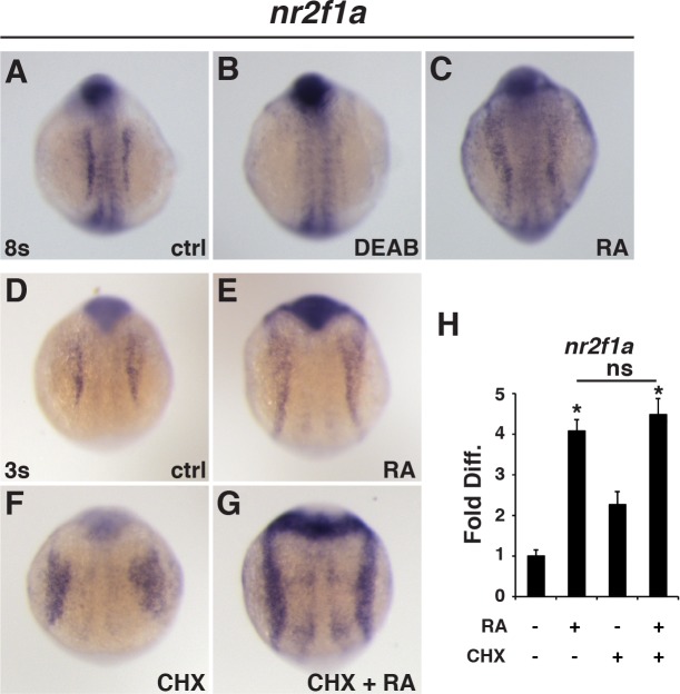
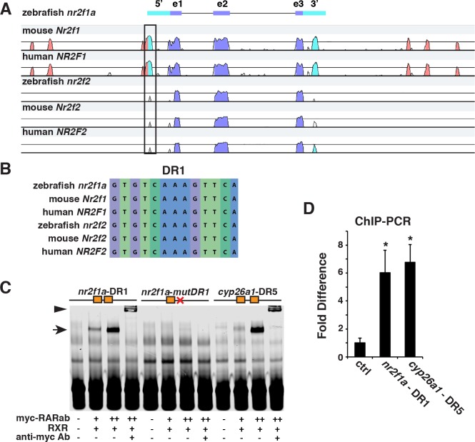
RA responsiveness of NR2F genes is conserved in chordates [28, 42–44]. We identified nr2f1a as an RA-responsive gene within the ALPM of zebrafish embryos (Fig 1A–1C), consistent with what other groups have described [28, 44]. However, the nature of this regulation has not been assessed. Furthermore, although RA signaling affects epigenetic modifiers that control the expression of Nr2f1 in mammalian cells, a direct role for RA signaling has not been shown [30]. We found that RA treatment positively regulates nr2f1a expression after cycloheximide (CHX) treatment (Fig 1D–1H), implicating a direct transcriptional regulatory mechanism. To determine if there are putative RA response elements (RAREs) for RAR binding sites in the nr2f1a promoter region, we first performed a mVISTA alignment of zebrafish, mouse, and human NR2F1 and NR2F2 genomic sequences. We found a highly conserved region within the 5’-untranslated region (UTR) of nr2f1a (Fig 2A). Using the nuclear hormone receptor binding site prediction tool NHRscan in this region, we found a completely conserved direct repeat 1 (DR1) site [45–49] within the 5’-UTR of these genes (Fig 2B). While the location of this DR1 site is atypical, regulatory elements of other genes have been found to overlap with the 5’-UTR [50, 51]. Despite the conservation of these sites across phyla, the site was not present in the zebrafish paralog nr2f1b, which is not RA responsive [28]. Electrophoretic mobility shift assays (EMSAs) and chromatin immunoprecipitation-quantitative PCR (ChIP-qPCR) indicated that RARs can bind the nr2f1a DR1 in vitro and in vivo (Fig 2C and 2D). However, this site was not sufficient to respond to RA alone in luciferase assays (S1 Fig). Therefore, our results suggest RA directly regulates nr2f1a expression and may involve interactions with a conserved DR1 RARE, although this atypical site may not be responsive to RA through a canonical activation mechanism.
Fig 1. RA signaling directly regulates nr2f1a expression.
(A-C) In situ hybridization (ISH) for nr2f1a in control (ctrl), DEAB-treated, and RA-treated embryos at the 8s stage. (D-G) ISH for nr2f1a in the LPM of ctrl, RA, CHX, and RA+CHX treated embryos. (H) RT-qPCR of nr2f1a expression after RA and CHX treatments at the 3s stage. Asterisks in all graphs indicate statistically significant difference from controls with p<0.05.
Fig 2. RARs bind a conserved DR1 site within the nr2f1a promoter.
(A) mVISTA sequence alignment of 17kb coding and flanking regions for zebrafish, mouse, and human NR2F1 and NR2F2 genes. Box indicates region containing the conserved DR1 site. purple–conserved coding sequence, blue–conserved 5’- and 3’-UTRs, red–conserved sequences outside of the transcribed regions. (B) Conserved DR1 sequence in NR2F genes. (C) EMSA using probes for the nr2f1a - DR1, a mutated nr2f1a - DR1, and control cyp26a1—DR5 sites with increasing amounts of myc-tagged RARabv2 protein. Zebrafish RXRba protein was added to all samples as binding was not observed without RXR. (D) ChIP-qPCR of the nr2f1a DR1 site, negative control site, and a previously reported Cyp26a1 DR5 site (positive control) comparing the association of induced VP16-RARab from hsp70l:EGFP-VP16-RARab embryos to that in non-transgenic control sibling embryos at the 8s stage.
Nr2f1a and nr2f2 are redundantly required to restrict ventricular CM and promote posterior PM development
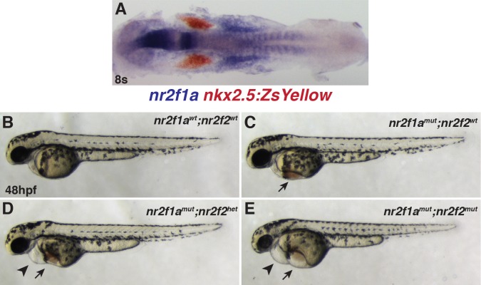
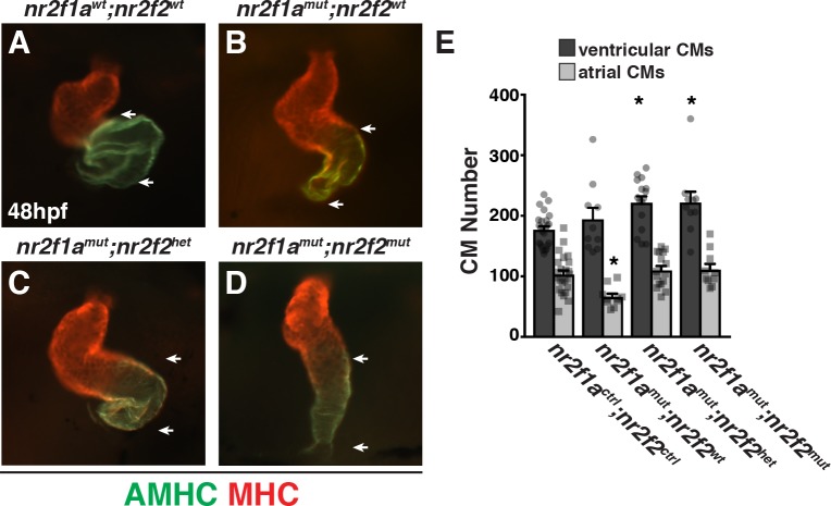
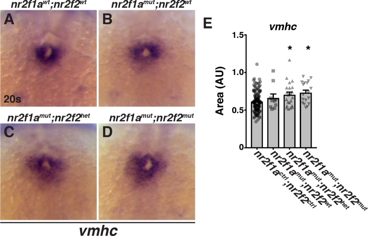
Within the ALPM, zebrafish nr2f1a is expressed immediately posterior to cardiac progenitors during somitogenesis (Fig 3A). However, our recent study of nr2f1a mutants did not reveal requirements for Nr2f1a at these early developmental stages when the cardiac progenitor field is established [39]. Instead, we found that Nr2f1a is required to promote atrial CM differentiation at both the arterial and venous poles of the atrial chamber at subsequent stages of cardiogenesis, consistent with its expression specifically in atrial CMs within the developing cardiac tube [39]. Although zebrafish nr2f2 mutants do not have overt cardiovascular defects through at least two weeks of development [37, 38], zebrafish nr2f2 has low levels of expression within the ALPM during somitogenesis and is responsive to RA signaling (S2 Fig), albeit significantly less so than nr2f1a as has been previously shown [28]. Therefore, we wondered if Nr2f2 functions redundantly with Nr2f1a at earlier stages of development within the ALPM. Using established engineered zebrafish nr2f2 deletion mutants [38], we found that loss of either one or both wild-type (WT) nr2f2 alleles in nr2f1a mutant embryos resulted in overall progressively worse pericardial and yolk edemas coupled with blood pooling on the yolk compared to nr2f1a mutants alone (Fig 3B–3E). Similarly, we found that loss of nr2f2 alleles in nr2f1a mutants produced hearts that were more dysmorphic and linear than nr2f1a mutant hearts alone (Fig 4A–4D). Despite the exacerbation of the cardiac dysmorphology in the compound nr2f1a; nr2f2 mutants, we did not observe enhanced reduction of atrial chamber size or expression of AMHC, a marker of differentiated atrial CMs (Fig 4A–4D). Valve markers were also not further expanded with the loss of nr2f2 alleles in nr2f1a mutants (S3 Fig), consistent with a unique role of Nr2f1a in limiting valve development [39]. Surprisingly, in contrast to nr2f1a mutants, which display a specific reduction in atrial CMs (Fig 4E; [39]), counting CMs with the myl7:h2afva-mCherry transgene [52] revealed that loss of one or both nr2f2 alleles in nr2f1a mutants produced an equivalent increase in ventricular CMs without producing any deficit in atrial CMs (Fig 4E). Although we have found that the loss of atrial CMs is not due to early specification defects within the ALPM of nr2f1a mutants [39], we posited that the specific surplus of ventricular CMs in the nr2f1a; nr2f2 mutants is due to an increase in ventricular CM specification at earlier stages of cardiogenesis because both nr2f1a and nr2f2 are expressed within in the ALPM [28]. Consistent with this idea, in the double mutants we observed a modest expansion of the cardiac progenitor marker Nkx2.5 at the 16 somite (s) stage (S4 Fig) and the amount of differentiating ventricular CMs, indicated by ventricular myosin heavy chain (vmhc), was increased at the 20s stage (Fig 5A–5E). Furthermore, loss of both nr2f1a and nr2f2 appeared to partially repress the ability of RA to inhibit vmhc expression (S5 Fig). Together, these data suggest that Nr2f1a and Nr2f2 function redundantly to restrict the number of differentiating ventricular CMs.
Fig 3. Loss of nr2f2 alleles in nr2f1a mutants produces stronger overt cardiovascular defects.
(A) Two-color ISH for nr2f1a (blue) and nkx2.5:ZsYellow (red) at the 8s stage. Embryo is flat-mounted with dorsal view and anterior left. (B-E) Lateral views of the nr2f1awt; nr2f2wt, nr2f1amut; nr2f2wt, nr2f1amut; nr2f2het, and nr2f1amut; nr2f2mut embryos. n = 16 overtly WT embryos and n = 32 embryos with edema that were genotyped for the experiment shown. While embryos that have two WT alleles are shown, the other combinations of nr2f1a and nr2f2 alleles, other than nr2f1ahet; nr2f2mut embryos, were indistinguishable from WT embryos at 48 hpf. 1 of 5 embryos that genotyped as nr2f1ahet; nr2f2mut embryos was indistinguishable from WT, while 4 of the 5 nr2f1ahet; nr2f2mut embryos displayed a very small amount of blood pooling on the yolk. However, cardiac defects were not found in these embryos. Arrows indicate edema and blood pooling on the yolk. Arrowheads indicated pericardial edema.
Fig 4. Nr2f1a functions redundantly with Nr2f2 to restrict ventricular CM number.
(A-D) Frontal view of hearts from nr2f1awt; nr2f2wt, nr2f1amut; nr2f2wt, nr2f1amut; nr2f2het, and nr2f1amut; nr2f2mut embryos at 48 hpf with immunohistochemistry (IHC). Atria (AMHC)—green. Ventricles (MHC)—red. (E) CM number from hearts of nr2f1actrl; nr2f2ctrl (n = 24), nr2f1amut; nr2f2wt (n = 10), and nr2f1amut; nr2f2het (n = 16), and nr2f1amut; nr2f2mut (n = 10) embryos with the myl7:h2afva-mCherry transgene at 48 hpf. Although embryos WT for nr2f1a and nr2f2 alleles are shown in all images for comparison to nr2f1a; nr2f2 mutants, other allele combinations were indistinguishable from WT and pooled for data analysis. Therefore, nr2f1actrl; nr2f2ctrl indicates analysis performed with the combination of nr2f1awt; nr2f2wt; nr2f1ahet; nr2f2wt, nr2f1awt; nr2f2het, and nr2f1ahet; nr2f2het embryos.
Fig 5. Nr2f1a and Nr2f2 redundantly restrict differentiation of ventricular CMs.
(A-D) ISH for vmhc in nr2f1awt; nr2f2wt, nr2f1amut; nr2f2wt, nr2f1amut; nr2f2het, and nr2f1amut; nr2f2mut embryos at the 20s stage. (E) Area measurements in arbitrary units (AU) of vmhc expression from nr2f1actrl; nr2f2ctrl (n = 141), nr2f1amut; nr2f2wt (n = 8), nr2f1amut; nr2f2het (n = 22), and nr2f1amut; nr2f2mut (n = 18) embryos at the 20s stage.
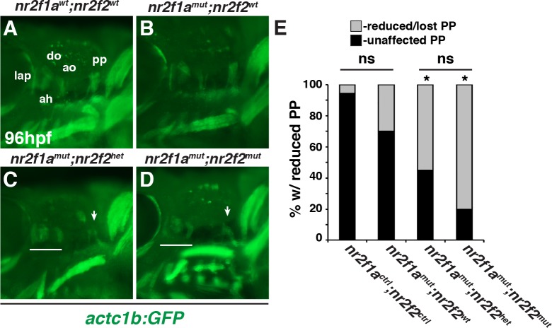
Previous analysis suggested that loss of RA signaling does not promote an increase in cardiac progenitor proliferation within the ALPM [53]. Consistent with this data, we did not find an increase in the number of proliferating Nkx2.5+ cells at the 16s stage in nr2f1a; nr2f2 mutant embryos (S4 Fig). Thus, we postulated that the surplus ventricular CM progenitors in nr2f1a; nr2f2 mutant embryos, which refers to nr2f1amut with either nr2f2het or nr2f2mut alleles, may be at the expense of an adjacent cell lineage. We reasoned that candidates were the pharyngeal arch arteries (PAAs) and PMs, since their progenitors intermingle with the cardiac progenitor population within the anterior mesoderm of zebrafish [5, 54]. We examined the posterior PAAs and PMs in nr2f1a; nr2f2 mutants at 48 hpf and 96 hpf, developmental time points when these cells have respectively differentiated [55]. Interestingly, we did not detect defects in PAA number and morphology in nr2f1a; nr2f2 mutant embryos carrying the kdrl:EGFP transgene (S6 Fig). However, in contrast to the PAAs, we found the posterior protractor pectoralis (pp), which is proposed to be a homolog of vertebrate neck muscles derived from the occipital LPM [56–60], was often lost or reduced in nr2f1a; nr2f2 mutant embryos (Fig 6A–6E). Although not as dramatic, the anterior dorsal mandibular (1st) and hyoid (2nd) arch derived muscles were also often smaller and disorganized compared to WT and nr2f1a mutant siblings (Fig 6A–6D). A similar trend with respect to increased pp loss was observed at 75 hpf (S7 Fig). However, for the analysis of the compound mutants we focused on 96 hpf to ensure that any defects were not due to developmental delay. Together, these data suggest that Nr2f1a and Nr2f2 together are required to promote posterior PM development.
Fig 6. Nr2f1a; nr2f2 mutants have deficiencies in their posterior PMs.
(A-D) PMs in nr2f1awt; nr2f2wt, nr2f1amut; nr2f2wt, nr2f1amut; nr2f2het, and nr2f1amut; nr2f2mut embryos with the actc1b:GFP transgene at 96 hpf. IHC was performed for GFP. Views are lateral with anterior to the left and dorsal up. Arrows in C and D indicate largely absent protractor pectoralis (pp). Brackets in C and D indicate 1st and 2nd arch PMs. ah–adductor hyoideus, ao–adductor operculi, do–dilator operculi, lap–levator arcus palitini. Muscle nomenclature used is from [55]. (E) Percentage of nr2f1actrl; nr2f2ctrl (n = 18), nr2f1amut; nr2f2wt (n = 10), nr2f1amut; nr2f2het (n = 29), and nr2f1amut;nr2f2mut (n = 15) embryos with loss or malformed pp muscles at 96 hpf.
Lineage tracing of tcf21+-derived progeny in nr2f1a; nr2f2 mutant embryos
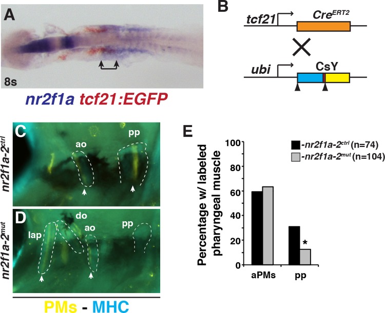
Due to the inverse effects on ventricular CM and posterior PM development in the nr2f1a; nr2f2 mutants, we sought to understand the relationship of these progenitors. Using two-color ISH to examine the expression of nr2f1a relative to tbx1 and tcf21, we found that nr2f1a expression does not significantly overlap with tbx1 (S8 Fig). However, nr2f1a and tcf21 expression domains overlap in a caudal region of the ALPM (Fig 7A), interestingly, where lineage tracing has shown tcf21+ progeny give rise to CMs and posterior PM [5]. Despite the overlap in expression, tcf21 expression was not affected in nr2f1a; nr2f2 mutant embryos (S8 Fig). Since the tcf21+ progenitors are overtly specified properly in nr2f1a; nr2f2 mutant embryos, we hypothesized that Nr2f proteins may affect a fate decision of progenitors within the posterior ALPM that can become ventricular and/or PM progenitors. To test this, we first used the inducible tcf21:CreERT2 transgene with the Cre-mediated color-switch line ubi:LOXP-AmCyan-STOP-LOXP-ZsYellow (CsY) to permanently label cells that have expressed tcf21+ (Fig 7B). For lineage tracing experiments, nr2f1a homozygous mutants (nr2f1amut) coupled with nr2f2 heterozygosity (nr2f2het) or nr2f2 mutant homozygosity (nr2f2mut) were analyzed together (referred to as nr2f1a-2mut), because our data suggest loss of a single WT nr2f2 allele in nr2f1a mutants produces similar ventricular CM and PM defects as loss of both WT alleles in nr2f1a mutants. Consistent with what has been reported [5], we found that tamoxifen treatment of embryos containing both transgenes produced labeling of skeletal muscle within the PMs (Fig 7C–7E). Although we did not find a decrease in the frequency of labeled anterior PMs within the 1st and 2nd arches, we found a decrease in the frequency of contribution to the pp in the nr2f1a-2mut embryos (Fig 7E), supporting that Nr2f proteins promote the differentiation of skeletal muscle within the pp.
Fig 7. Tcf21+ progenitors less frequently contribute to the pp muscle in nr2f1a-2 mutant embryos.
(A) Two-color ISH of nr2f1a (blue) and tcf21:EGFP (red). Embryo is flat-mounted with anterior leftward. Bracket indicates region of overlap. (B) Schematic of tcf21:CreERT2 recombinase and ubiquitous Cre-mediated color-switch transgenic lines used. (C, D) PMs (arrowhead) labeled in nr2f1a-2ctrl and nr2f1a-2mut embryos with the tcf21:CreERT2; ubi:CsY transgenes. Labeled PMs–yellow. Muscles (MHC)–blue. Outlines indicate PMs with labeled skeletal muscles. While other cells were labeled within the pharyngeal region, they were not scored as skeletal muscle because they were not located within the muscles or had morphology consistent with skeletal muscle. View is lateral with anterior to the left and dorsal up. (E) Percentage of labeled PMs on each side of the nr2f1a-2ctrl (n = 74) and nr2f1a-2mut (n = 104) embryos. The (n) reflects the total number of sides examined, since labeling was not equivalent on both side of an embryo. aPMs—anterior pharyngeal muscles, pp—protractor pectoralis. 44/74 nr2f1a-2ctrl and 66/104 nr2f1a-2mut had muscle labeled in aPMs. 23/74 nr2f1a-2ctrl and 13/104 nr2f1a-2mut had muscle labeled in the pp. Fisher’s exact test was used to determine if there was a difference between the frequency of anterior and posterior PMs in nr2f1a-2ctrl and nr2f1a-2mut embryos.
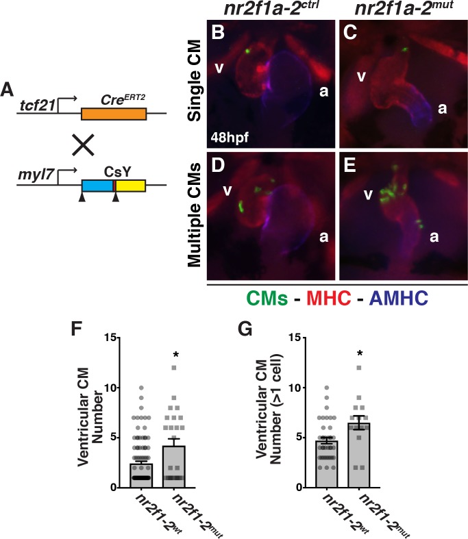
We then reasoned that if Nr2f proteins are influencing a fate decision of ventricular and PM progenitors, tcf21+ progenitors should become ventricular CMs at an increased frequency in nr2f1a; nr2f2 mutant embryos. While we found that using tcf21:CreERT2; ubi:CsY labeled a few CMs, the expression was not as robust as for the PM. Therefore, we used the myl7:CsY transgene in combination with the tcf21:CreERT2 transgene to specifically and permanently label CMs derived from tcf21+ progenitors (Fig 8A–8E). Examining labeled ventricular CMs, we found a trend where nr2f1a-2mut embryos have an increase in the number of embryos with >1 tcf21+-derived ventricular CM labeled compared to control embryos (S9 Fig). Importantly, overall, nr2f1a-2mut embryos on average have about twice as many tcf21+-derived ventricular CMs compared to WT sibling embryos (Fig 8F). Furthermore, there were increased number of labeled ventricular CMs found in nr2f1a-2mut embryos when just examining the pool of embryos that had >1 ventricular CM labeled (Fig 8G), further supporting an increase in the frequency and number of tcf21+-derived ventricular CMs contributing to the ventricles in nr2f1a-2mut embryos. While atrial CMs were also labeled, their labeling was infrequent compared to labeling of ventricular CMs (S9 Fig). We did not find a statistical difference in the frequency or average number of atrial CMs labeled within the populations (S9 Fig). Together, our lineage tracing of tcf21+ progenitors demonstrates that a greater number of their progeny give rise to ventricular CMs in nr2f1a-2mut embryos, while fewer give rise to the pp.
Fig 8. Nr2f proteins are required to limit ventricular CMs.
(A) Schematic of tcf21:CreERT2 recombinase and myl7 promoter driven Cre-mediated color-switch transgenic lines used. (B-E) Hearts with labeled CMs in nr2f1a-2ctrl and nr2f1a-2mut embryos carrying the tcf21:CreERT2; myl7:CsY transgenes. Labeled CMs–green. Cardiac muscles (MHC)–red. Atrium (AMHC)–blue. Images are frontal views. (F) Mean number of ventricular CMs labeled in nr2f1a-2wt (n = 98) and nr2f1a-2mut (n = 24) embryos. (G) Mean number of ventricular CMs labeled when more than one labeled ventricular CM was found in nr2f1a-2wt (n = 38) and nr2f1a-2mut (n = 14) embryos.
Discussion
Previous studies have demonstrated that RA signaling is necessary to limit cardiac specification and promote PM development [22, 61]. With respect to heart development, early RA signaling restricts the posterior border of atrial and ventricular progenitors within the ALPM [25]. Despite similar effects on both cardiac cell types, mechanisms restricting atrial CMs and ventricular CMs downstream of RA signaling appear to be temporally distinct [25]. The present study suggests that Nr2f1a and Nr2f2 function redundantly downstream of RA signaling within the ALPM to regulate these converse effects on ventricular CM and PM specification. While there are numerous similarities between our observations in comparison to RA signaling-deficient embryos [22, 25], it is worth recognizing that the heart and PM defects found in nr2f1a; nr2f2 mutant embryos are less severe than what is typically found with RA signaling-deficient embryos. Therefore, we hypothesize that these Nr2f transcription factors likely are part of a larger RA-responsive gene network, including Hox genes and Fgf signaling, that contributes to this allocation of progenitors within the ALPM.
Recent work examining Nr2f proteins in cranial neural crest that generate the anterior jaw has suggested significant redundancy with Nr2f1b and Nr2f5 in that developmental context [38]. However, we have not found any evidence of redundancy or genetic interactions with Nr2f1b and/or Nr2f5 in regulating heart development. For example, unlike what is observed with nr2f1a and nr2f2, nr2f1a+/-;nr2f5+/- intercrosses produce ~25% (16/56) mutant embryos that are indistinguishable from nr2f1a mutant embryos with respect to the heart and blood pooling. We also have not found evidence for compensatory expression of any nr2f genes in the nr2f1a mutants (S10 Fig). Additionally, we have not found defects in neural crest markers in the nr2f1a; nr2f2 mutants (S8 Fig), suggesting loss of the pp is not secondary to neural crest defects. Our recent work suggests that Nr2f1a alone functions to promote atrial CM differentiation as the heart elongates and atrial CMs mature [39], which is after it first appears in the ALPM. Here, we demonstrate that the change in the number of atrial CMs is not exacerbated in the nr2f1a; nr2f2 mutant embryos compared to nr2f1a single mutants. Instead, the number of atrial CMs is increased relative to nr2f1a single mutants, despite a similar overt reduction in atrial chamber size and lack of AVC, and equivalent to the number found in control embryos. We posit that these differential effects on the production of atrial CMs are because Nr2f proteins restrict the posterior extent of both atrial and ventricular progenitor fields within the ALPM and that a deficit in differentiating atrial CMs is not observed because the earlier requirements limiting the cardiac progenitor field offset the later requirements promoting atrial CM differentiation.
NR2Fs are conserved regulators of atrial chamber development in vertebrates. Zebrafish nr2f1a mutants and mouse global Nr2f2 KOs present smaller atria [35, 39], while conditional Nr2f2 KO in the heart at later stages suggests a role in maintenance of atrial CM identity [36]. NR2F2 is required for atrial CM differentiation in human iPSCs [32]. Given these conserved requirements, it is interesting to compare the phenotypes of the nr2f1a; nr2f2 double mutants to the variability and severity of CHDs associated with NR2F2 lesions in humans. It has been proposed there is a direct correlation between the severity of CHDs and types of lesions impacting NR2F2 function [41]. Specifically, nonsense mutations proposed to be more damaging and resulting in significant loss of NR2F2 predominantly are associated with LVOTO, while missense mutations proposed to be less damaging are associated with ASDs and AVSDs [41]. The variable CHDs affecting the arterial pole of the ventricle and the atrial chamber are highly reminiscent of the chamber-specific defects we observe in nr2f1a; nr2f2 double mutants compared to single nr2f1a mutants, which overtly affect the production of ventricular CMs and atrial CMs, respectively. Together, these data support the hypothesis that levels of total Nr2f dosage differentially affect chamber-specific cardiogenic processes within the vertebrate heart. Moreover, we propose that greater loss of NR2F transcription factors, through more damaging alleles or genetic loss, produces ventricular chamber defects due to earlier developmental requirements within the ALPM, while ASDs or AVSDs may occur due to a more modest loss of total NR2F signaling that is required at later stages of atrial CM differentiation. Thus, our studies offer a working model to explain the molecular etiology of congenital LVOTO and ASDs/AVSDs associated with NR2F2 mutations in humans.
While NR2F proteins have been studied in numerous development contexts, significant analysis of the requirements for Nr2fs in skeletal muscle have not been reported. Virtually all the Nr2f proteins are expressed in the somites of zebrafish [28]. Interestingly, in mice Nr2f2 is broadly expressed in skeletal muscle, including the somites and the cranial muscles [34, 41]. Limb-specific Nr2f2 KOs indicate it is required for limb muscle development [34] and mechanistically there is evidence that Nr2f2 can compete with myoD in muscle differentiation [62]. Therefore, there is precedence for Nr2f2 functions in somite-derived skeletal muscle, but requirements in PM development have not been reported. It is interesting to note that cranio-facial defects have been associated with genetic deficiencies that affect both NR2F1 and NR2F2 in humans [40, 63, 64]. In two independent cases, similar-sized deletions that eliminate NR2F2 were associated with cranial abnormalities as well as ASDs [40, 64]. However, overt cranio-facial defects similar to those found in the deficiencies were not reported in patients found to have specific mutations that affect NR2F2 and are associated with CHDs [41]. Therefore, although specific defects in craniofacial muscle were not reported, there is precedence for an association between NR2F gene loss and both craniofacial and CHDs in humans.
Recent clonal analysis in mice has suggested there are common cardio-pharyngeal progenitors that contribute progeny to the neck muscles, the arterial pole and atria that are distinct from other cardio-pharyngeal populations of the SHF [8]. Our data are also consistent with a close association of ventricular outflow tract and pharyngeal neck muscle progenitors and a distinction from other SHF progenitors, which arise more anteriorly [65, 66]. Specifically, while anterior dorsal 1st and 2nd arch muscles are reduced, we predominantly find that nr2f1a; nr2f2 embryos lose the posterior pp muscle (cucullaris), which has been proposed to be homologous to the ALPM-derived trapezius neck muscles in mammals [56–60]. Therefore, reminiscent of the recent retrospective clonal analysis in mice [8], these results hint at the existence of a distinct posterior progenitor population with cardiac and PM potential that does not correspond to the anterior SHF.
Given the existence of bi-potent cardio-pharyngeal progenitors in mice and Ciona [6–8, 19, 20], one interpretation of our results is that RA signaling and consequently Nr2f proteins, at least in part, act on bi-potent cardio-pharyngeal progenitors. Although it is clear from the retrospective clonal lineage analysis in mice that there are multiple populations of bi-potent cardio-pharyngeal progenitors, these populations are rare and only found from examination of large sample sizes [6–8]. While zebrafish tcf21+, as well as nkx2.5+ progenitors, can give rise to ventricular CMs and PMs [5, 67], it is not yet clear whether there are bi-potent progenitors with cardiac and PM potential. Although the defects we observe in ventricular CM and PM development of nr2f1a; nr2f2 mutant embryos are less dramatic than with loss of RA signaling, in neither case are the defects subtle enough that a very rare population of bi-potent progenitors is likely being affected. Instead, we favor a model where there is a larger population of progenitors within the ALPM that have the potential to become either ventricular CMs and PM, with signals such as Nr2f proteins functioning downstream of RA signaling to influence their allocation into one of these populations.
Overall, our study provides valuable insight into the requirements of Nr2f genes in vertebrate cardiac and cranial muscle development. These studies may help us to further understand molecular and genetic etiology controlling phenotypic variability of CHDs as well as developmental syndromes that have congenital malformations concomitantly affecting the heart, head, and neck muscles in humans.
Methods
Ethics statement
All zebrafish husbandry and experiments were performed in accordance with protocols approved by the Institutional Animal Care and Use Committee (IACUC) of Cincinnati Children's Hospital Medical Center.
Zebrafish line and maintenance
Adult zebrafish were raised and maintained under standard laboratory conditions. Transgenic lines used were: Tg(kdrl:nlsEGFP)ubs1 [68], Tg(kdrl:EGFP)la116, TgBAC(−36nkx2.5:ZsYellow)fb7 [69], Tg(actc1b:GFP)zf13 [70], Tg(tcf21:nucEGFP)pd41 [71], Tg(tcf21:CreERT2)pd42 [72], Tg(ubi:LOXP-AmCyan-STOP-LOXP-ZsYellow)fb5 [69], Tg(myl7: LOXP-AmCyan-STOP-LOXP-ZsYellow)fb2 [69], Tg(myl7:h2afva-mCherry)sd12 [52] and Tg(hsp70l:EGFP-VP16-RAR-)c1004 [73]. Mutant alleles used were: nr2f1ael512 and nr2f2el506 [38, 39].
Whole mount ISH and area measurements
Single and two-color whole mount ISH were performed using NBT/BCIP (Roche) and INT/BCIP (Roche), as previously reported [74]. Digoxygenin- and fluorescein-labeled anti-sense RNA probes for zsyellow (ZDB-EFG-110824-1), egfp (ZDB-EFG-070117-1), nr2f1a (ZDB-GENE-980526-115), nr2f2 (ZDB-GENE-990415-252), vmhc (ZDB-GENE-991123-5), nkx2.5 (ZDB-GENE-980526-321), dlx2a (ZDB-GENE-980526-212), tbx1 (ZDB-GENE-030805-5), tcf21(ZDB-GENE-051113-88), and klf2a (ZDB-GENE-011109-1) were used. Area measurements were performed using ImageJ.
Analysis of Nr2f loci and promoters
Sequences for zebrafish, mouse, and human Nr2f genes plus a 10kb region 5’ and 3’ to the genes were taken from Ensembl (ensembl.org) and aligned using mVista (http://genome.lbl.gov/vista/mvista/submit.shtml). Locations, excluding exons, in which there was over 50% conservation between any of the sequences were analyzed for the presence of RARs. Conserved sequences were input into NHRscan (http://www.cisreg.ca/cgi-bin/NHR-scan/nhr_scan.cgi) to identify potential RAR binding sites.
RT-qPCR
Total RNA isolation and RT-qPCR was performed using previously reported methods [75]. Briefly, whole embryo RNA was obtained from groups of 30 embryos using Trizol (Ambion) and Purelink RNA Microkit (Invitrogen). cDNA was synthesized using 1μg total RNA and the ThermoScript Reverse Transcriptase kit (Invitrogen). RT-qPCR was performed using Power SYBR Green PCR Master Mix (Applied Biosystems) in a BioRad CFX-96 PCR machine. Expression levels were standardized to β-actin expression and data were analyzed using the 2-ΔΔCT Livak Method. All experiments were performed in triplicate. Primer sequences for β-actin were reported previously [73, 75]. All primer sequences used for RT-qPCR are in the S1 Table.
Drug treatments
All drug treatments were administered to embryos in 2 mL of blue water with drug at specified concentrations in a glass vial with 25–30 embryos/vial at 28.5°C. For analysis of nr2f1a and nr2f2 expression, embryos were treated with CHX (10 μM, Sigma 48591), RA (1 μM, Sigma R2625), and DEAB (10 μM, Sigma D86256) at tailbud stage for 1 hour. For analysis of vmhc expression in nr2f1a; nr2f2 mutants, embryos were treated with 0.05 μM RA at the 3s stage until the 20s stage. Drugs were washed out 3X with embryo water then the embryos were fixed in 4% formaldehyde for analysis. Vmhc-stained embryos were genotyped following imaging. Tamoxifen (10 μM, Sigma H7904) was administered in 30 mL of blue water with 0.003% PTU in petri dishes to embryos at 30%-50% epiboly until embryos were analyzed or through 2 days of development.
ChIP-qPCR
ChIP-qPCR was performed essentially as previously reported [75]. Hemizygous Tg(hsp70l:VP16-RAR:EGFP)c1004 adults were crossed to WT adult zebrafish. The resulting embryos were collected at tailbud stage and heat-shocked at 37°C for 30 minutes. Transgenic embryos were sorted from their non-transgenic control siblings by the presence of GFP. Embryos (n = 100) were dechorionated and fixed in 1% formaldehyde 2 hours after heat-shock. Cells were lysed by gentle pipetting in cell lysis buffer. Nuclei were lysed and DNA was sheered by sonication with glass beads to 200-600bp fragments. Dynabeads (Invitrogen) were used to pull down GFP tagged proteins with ChIP-grade polyclonal anti-GFP antibody (Abcam ab290) per manufactures instructions. Samples were de-crosslinked and qPCR was used to quantify the fold difference in enrichment of the DR1 RARE in the nr2f1a promoter and the known DR5 RARE in the Cyp26a1 promoter as compared to a nr2f1a promoter region not containing a RARE. Expression levels were standardized to the no antibody control signal and data were analyzed using the 2-ΔΔCT Livak Method. Primer sequences for cyp26a1 ChIP-PCR were reported previously [76]. Primer sequences for nr2f1a DR1 ChIP-PCR and control are indicated in S1 Table.
EMSA
EMSA was performed essentially as previously reported [77]. Oligonucleotides were designed containing the nr2f1a DR1 site (GTGTCAAAGTTCA), the nr2f1a DR1 site with a targeted mutation in the second half site of the DR1 abolishing the direct repeat (GTGTCAAAGTCAT), and a previously reported Cyp26a1 DR5 site [76]. A complementary oligonucleotide was designed with a 5’ LI-COR IRDye 700 (IDT). The oligonucleotides were annealed and the ends filled with Klenow (New England Biolabs). Zebrafish myc-rarab was in the pCS2+MT. Zebrafish RXRba was cloned into pCS2p+. Proteins for EMSA were made using the TnT SP6 Quick Coupled Transcription/Translation System (Promega). Protein samples were gently mixed with LI-COR tagged probes and incubated at room temperature for 20 minutes. 4% polyacrylamide gels were run for 2 hours at 150 V. Gels were imaged using an Odyssey CLx LI-COR imager.
Zebrafish IHC and CM counts
Embryos were fixed for 1 hour at room temperature in 1% formaldehyde in PBS in 3 ml glass vials. Embryos were washed 1X in PBS and then 2X in 0.2% saponin/1X PBS, followed by blocking in 0.2% saponin/0.5%sheep serum/1X PBS (Saponin blocking solution) for one hour. AMHC (S46) and MHC (sarcomeric myosin; MF20) primary antibodies (Developmental Studies Hybridoma Bank) were incubated at 1:10 in Saponin blocking solution. Rabbit polyclonal DsRed antibody (Clontech), to detect mCherry, and Living colors anti-RCFP (Clontech), to detect ZsYellow, were used at a 1:1000 dilution. Rabbit anti-GFP (Abcam) was used at 1:500. Rabbit anti-Nkx2.5 (Gene Tex) was used at 1:250. Mouse anti-pHH3 (Abcam) was used at 1:1000. All secondary antibodies were used at dilutions of 1:100. Antibody information is also listed in S2 Table. Cell counts were performed by gently flattening embryos under a coverslip and counting the fluorescent nuclei in each chamber. For all imaging except Nkx2.5/pHH3, embryos were imaged using a Zeiss M2BioV12 Stereo microscope. For Nkx2.5/pHH3, embryos were post-fixed in 2% formaldehyde/1X PBS for two hours and mounted in 1% low-melt agar on 2% agar plates. Images of one side of the embryo were taken using a Nikon A1R Multiphoton Upright Confocal Microscope with a 16X water immersion objective. 200 μm optical sections were taken with the resonance scanner.
Luciferase assays
The promoter fragments for both reporters used were cloned into the Kpn and HindIII sites of the pGL3 (Promega) multiple cloning site. The DR1-ef1a construct contains 165 base pairs (bp) of the nr2f1a promoter and 5’UTR adjacent to 193 bp of a minimal elongation factor 1a (ef1a) promoter (green). The nr2f1a-DR1 construct contains 371 bp that include the promoter and 5’UTR containing the conserved DR1 site. The pGL3-12XRARE-tk vector and dual luciferase assays were reported and performed in HEK293 cells as described previously [78].
Statistical analysis
To compare two groups, we performed a Student’s t-test or Mann-Whitney test. To compare 3 or more conditions are different, we performed ANOVA analysis. To determine if two proportions were statistically distinct we performed a Chi-squared test or Fisher’s exact test. Statistical analysis was performed using GraphPad Prism. A p value < 0.05 was considered statistically significant for all analysis.
Supporting information
(A) Schematic of the two constructs placed into pGL3. The pGL3-DR1-ef1a construct has nucleotides -60 of the nr2f1a promoter (blue) through +105 of 5’UTR (gray), which includes the including the DR1 site (yellow box) cloned adjacent to a minimal elongation factor 1a (ef1a) promoter (green) (358 bp). The pGL3-nr2f1a-DR1 construct contains nucleotides -266 through +105 (371 bp) of the promoter and 5’UTR containing the conserved DR1 site. Blue indicates nr2f1a promoter sequences. Red boxes indicate predicted TATA boxes. (B) Luciferase assays testing RA responsiveness in HEK 293 cells. FF–firefly luciferase. RL—renilla luciferase. The previously reported pGL3-12XRARE-tk plasmid [78] was used as a positive control.
(TIF)
(A-C) Nr2f2 expression in the ALPM of control, DEAB-treated, and RA-treated embryos. View is dorsal with anterior left. Arrows indicated anterior and posterior limits of expression in control and RA-treated embryos.
(TIF)
(A-D) ISH for the endocardial atrioventricular canal marker klf2a. Frontal views of hearts in nr2f1awt; nr2f2wt, nr2f1amut; nr2f2wt, nr2f1amut; nr2f2het, and nr2f1amut; nr2f2mut embryos. v–ventricle. a–atrium. Arrows indicate the length of klf2a expression within the hearts.
(TIF)
(A-D) ISH for the cardiac progenitor marker nkx2.5 in nr2f1awt; nr2f2wt, nr2f1amut; nr2f2wt, nr2f1amut; nr2f2het, and nr2f1amut; nr2f2mut embryos at the 16s stage. Dorsal view with anterior up. 160 embryos were examined with ≥9 embryos examined for each condition. Although we observed a trend in the expansion of nkx2.5 expression when assaying area of expression similar to vmhc, due to inherent variability in nkx2.5 expression and the low numbers of embryos, it was not statistically significant. (E,F) IHC for Nkx2.5 and pHH3 in nr2f1awt; nr2f2het and nr2f1amut; nr2f2mut embryos at the 16s stage. Confocal images of the ventro-lateral side of the embryo. Dorsal is right and anterior up. A single side of each embryo was used for analysis. (G) Number of Nkx2.5+ cells in control and nr2f1a; nr2f2 mutant embryos. (H) Percentage of pHH3+/Nkx2.5+ in control and nr2f1a; nr2f2 mutant embryos. For quantification of Nkx2.5+ and pHH3+/Nkx2.5+ cells, nr2f1a homozygous mutants (nr2f1amut) coupled with nr2f2 heterozygosity (nr2f2het) or nr2f2 mutant homozygosity (nr2f2mut) were analyzed together (referred to as nr2f1a-2mut), because our data suggest loss of a single WT nr2f2 allele in nr2f1a mutants produces a similar increase in ventricular CMs as double mutants. Nr2f1a-2ctrl includes any combination of nr2f1a and nr2f2 WT and heterozygous alleles. nr2f1a-2ctrl (n = 23) and nr2f1a-2mut (n = 9) for G and H.
(TIF)
(A-C) ISH for vmhc in control (untreated), RA-treated nr2f1awt; nr2f2het, and RA-treated nr2f1amut; nr2f2mut embryos at the 20s stage. Control embryos were not genotyped. (D) Percentage of embryos with the genotypes found that lacked vmhc expression (n = 16) or had vmhc expression (n = 16). Although a RA-treated nr2f1awt; nr2f2het is shown in B, nr2f1a-2ctrl includes any combination of nr2f1a and nr2f2 WT and heterozygous alleles. Fisher’s exact test was used to compare the frequency of embryos with two nr2f1amut alleles found in each condition.
(TIF)
(A,B) PAAs in nr2f1a-2ctrl and nr2f1a-2mut embryos. Numbers indicated arches. Anterior is to the right.
(TIF)
(A-D) PMs in nr2f1awt; nr2f2wt, nr2f1amut; nr2f2wt, nr2f1amut; nr2f2het, and nr2f1amut; nr2f2mut embryos at 75 hpf. Views are lateral with anterior to the left and dorsal up. (E) Percentage of nr2f1actrl; nr2f2ctrl (n = 7), nr2f1amut; nr2f2wt (n = 16), nr2f1amut; nr2f2het (n = 28), and nr2f1amut; nr2f2mut (n = 28) embryos with loss of posterior and malformed PMs at 75 hpf.
(TIF)
(A) ISH for tbx1 (red) and nr2f1a (blue) in the ALPM of an embryo at the 8s stage. Image is a dorsal view with anterior rightward of a flat-mounted embryo. (B-E) ISH for tcf21 in the ALPM of nr2f1awt; nr2f2wt, nr2f1amut; nr2f2wt, nr2f1amut; nr2f2het, and nr2f1amut; nr2f2mut embryos at the 18s stage. (F-I) ISH for the neural crest marker dlx2a in nr2f1awt; nr2f2wt, nr2f1amut; nr2f2wt, nr2f1amut; nr2f2het, and nr2f1amut; nr2f2mut embryos at the 18s stage. For B-I, views are dorsal with anterior up.
(TIF)
(A) Percentage of embryos with 1 and >1 ventricular CM. (B) Percentage of embryos with labeled CMs that had labeled atrial CMs. (C) Mean number of labeled atrial CMs in nr2f1a-2ctrl and nr2f1a-2mut embryos.
(TIF)
RT-qPCR for nr2f1b, nr2f2, nr2f5, nr2f6a, and nr2f6b in nr2f1a mutants at 48 hpf does not show compensatory expression.
(TIF)
(DOCX)
(DOCX)
Data Availability
All relevant data are within the manuscript and its Supporting Information files.
Funding Statement
This work was supported by grants from the National Institutes of Health (https://www.nih.gov): R01HL112893 and R01 HL137766 to J.S.W.; R01DE018405 to J.G.C.; K99DE026239 to L.A.B.; T32HL007382 to T.E.D. The funders had no role in the study design, data collection and analysis, decision to publish, or preparation of the manuscript.
References
- 1.Huxley JS, deBeer GR. The mosaic style of differentiation Barcroft J, Saunder JT, editors. London: Cambridge University Press; 1934. 195–270 p. [Google Scholar]
- 2.Jacobson AG, Sater AK. Features of embryonic induction. Development. 1988;104(3):341–59. . [DOI] [PubMed] [Google Scholar]
- 3.Tzahor E. Heart and craniofacial muscle development: a new developmental theme of distinct myogenic fields. Dev Biol. 2009;327(2):273–9. Epub 2009/01/24. doi: S0012-1606(09)00002-5 [pii] 10.1016/j.ydbio.2008.12.035 . [DOI] [PubMed] [Google Scholar]
- 4.Diogo R, Kelly RG, Christiaen L, Levine M, Ziermann JM, Molnar JL, et al. A new heart for a new head in vertebrate cardiopharyngeal evolution. Nature. 2015;520(7548):466–73. 10.1038/nature14435 . [DOI] [PMC free article] [PubMed] [Google Scholar]
- 5.Nagelberg D, Wang J, Su R, Torres-Vazquez J, Targoff KL, Poss KD, et al. Origin, Specification, and Plasticity of the Great Vessels of the Heart. Curr Biol. 2015;25(16):2099–110. 10.1016/j.cub.2015.06.076 [DOI] [PMC free article] [PubMed] [Google Scholar]
- 6.Lescroart F, Kelly RG, Le Garrec JF, Nicolas JF, Meilhac SM, Buckingham M. Clonal analysis reveals common lineage relationships between head muscles and second heart field derivatives in the mouse embryo. Development. 2010;137(19):3269–79. Epub 2010/09/09. 10.1242/dev.050674 . [DOI] [PubMed] [Google Scholar]
- 7.Lescroart F, Chabab S, Lin X, Rulands S, Paulissen C, Rodolosse A, et al. Early lineage restriction in temporally distinct populations of Mesp1 progenitors during mammalian heart development. Nat Cell Biol. 2014;16(9):829–40. 10.1038/ncb3024 . [DOI] [PMC free article] [PubMed] [Google Scholar]
- 8.Lescroart F, Hamou W, Francou A, Theveniau-Ruissy M, Kelly RG, Buckingham M. Clonal analysis reveals a common origin between nonsomite-derived neck muscles and heart myocardium. Proc Natl Acad Sci U S A. 2015;112(5):1446–51. Epub 2015/01/22. 10.1073/pnas.1424538112 [DOI] [PMC free article] [PubMed] [Google Scholar]
- 9.Abu-Issa R, Kirby ML. Heart field: from mesoderm to heart tube. Annu Rev Cell Dev Biol. 2007;23:45–68. Epub 2007/04/26. 10.1146/annurev.cellbio.23.090506.123331 . [DOI] [PubMed] [Google Scholar]
- 10.Dyer LA, Kirby ML. The role of secondary heart field in cardiac development. Dev Biol. 2009;336(2):137–44. 10.1016/j.ydbio.2009.10.009 [DOI] [PMC free article] [PubMed] [Google Scholar]
- 11.Guo C, Sun Y, Zhou B, Adam RM, Li X, Pu WT, et al. A Tbx1-Six1/Eya1-Fgf8 genetic pathway controls mammalian cardiovascular and craniofacial morphogenesis. J Clin Invest. 2011;121(4):1585–95. Epub 2011/03/03. 10.1172/JCI44630 [DOI] [PMC free article] [PubMed] [Google Scholar]
- 12.Harel I, Maezawa Y, Avraham R, Rinon A, Ma HY, Cross JW, et al. Pharyngeal mesoderm regulatory network controls cardiac and head muscle morphogenesis. Proc Natl Acad Sci USA. 2012;109(46):18839–44. Epub 2012/11/01. 10.1073/pnas.1208690109 [DOI] [PMC free article] [PubMed] [Google Scholar]
- 13.Felker A, Prummel KD, Merks AM, Mickoleit M, Brombacher EC, Huisken J, et al. Continuous addition of progenitors forms the cardiac ventricle in zebrafish. Nat Commun. 2018;9(1):2001 Epub 2018/05/23. 10.1038/s41467-018-04402-6 [DOI] [PMC free article] [PubMed] [Google Scholar]
- 14.Guner-Ataman B, Gonzalez-Rosa JM, Shah HN, Butty VL, Jeffrey S, Abrial M, et al. Failed Progenitor Specification Underlies the Cardiopharyngeal Phenotypes in a Zebrafish Model of 22q11.2 Deletion Syndrome. Cell reports. 2018;24(5):1342–54 e5. Epub 2018/08/02. 10.1016/j.celrep.2018.06.117 . [DOI] [PMC free article] [PubMed] [Google Scholar]
- 15.Baldini A. DiGeorge syndrome: the use of model organisms to dissect complex genetics. Hum Mol Genet. 2002;11(20):2363–9. Epub 2002/09/28. . [DOI] [PubMed] [Google Scholar]
- 16.Lu JR, Bassel-Duby R, Hawkins A, Chang P, Valdez R, Wu H, et al. Control of facial muscle development by MyoR and capsulin. Science. 2002;298(5602):2378–81. Epub 2002/12/21. 10.1126/science.1078273 . [DOI] [PubMed] [Google Scholar]
- 17.Nevis K, Obregon P, Walsh C, Guner-Ataman B, Burns CG, Burns CE. Tbx1 is required for second heart field proliferation in zebrafish. Dev Dyn. 2013;242(5):550–9. 10.1002/dvdy.23928 [DOI] [PMC free article] [PubMed] [Google Scholar]
- 18.Piotrowski T, Ahn DG, Schilling TF, Nair S, Ruvinsky I, Geisler R, et al. The zebrafish van gogh mutation disrupts tbx1, which is involved in the DiGeorge deletion syndrome in humans. Development. 2003;130(20):5043–52. 10.1242/dev.00704 . [DOI] [PubMed] [Google Scholar]
- 19.Stolfi A, Gainous TB, Young JJ, Mori A, Levine M, Christiaen L. Early chordate origins of the vertebrate second heart field. Science. 2010;329(5991):565–8. 10.1126/science.1190181 . [DOI] [PMC free article] [PubMed] [Google Scholar]
- 20.Wang W, Razy-Krajka F, Siu E, Ketcham A, Christiaen L. NK4 antagonizes Tbx1/10 to promote cardiac versus pharyngeal muscle fate in the ascidian second heart field. PLoS Biol. 2013;11(12):e1001725 Epub 2013/12/07. 10.1371/journal.pbio.1001725 [DOI] [PMC free article] [PubMed] [Google Scholar]
- 21.Niederreither K, Subbarayan V, Dolle P, Chambon P. Embryonic retinoic acid synthesis is essential for early mouse post-implantation development. Nat Genet. 1999;21(4):444–8. Epub 1999/04/07. 10.1038/7788 . [DOI] [PubMed] [Google Scholar]
- 22.Begemann G, Schilling TF, Rauch GJ, Geisler R, Ingham PW. The zebrafish neckless mutation reveals a requirement for raldh2 in mesodermal signals that pattern the hindbrain. Development. 2001;128(16):3081–94. . [DOI] [PubMed] [Google Scholar]
- 23.Keegan BR, Feldman JL, Begemann G, Ingham PW, Yelon D. Retinoic acid signaling restricts the cardiac progenitor pool. Science. 2005;307(5707):247–9. Epub 2005/01/18. 10.1126/science.1101573 . [DOI] [PubMed] [Google Scholar]
- 24.Sirbu IO, Zhao X, Duester G. Retinoic acid controls heart anteroposterior patterning by down-regulating Isl1 through the Fgf8 pathway. Dev Dyn. 2008;237(6):1627–35. 10.1002/dvdy.21570 . [DOI] [PMC free article] [PubMed] [Google Scholar]
- 25.Waxman JS, Keegan BR, Roberts RW, Poss KD, Yelon D. Hoxb5b acts downstream of retinoic acid signaling in the forelimb field to restrict heart field potential in zebrafish. Dev Cell. 2008;15(6):923–34. Epub 2008/12/17. 10.1016/j.devcel.2008.09.009 [DOI] [PMC free article] [PubMed] [Google Scholar]
- 26.Ryckebusch L, Wang Z, Bertrand N, Lin SC, Chi X, Schwartz R, et al. Retinoic acid deficiency alters second heart field formation. Proc Natl Acad Sci U S A. 2008;105(8):2913–8. doi: 0712344105 [pii] 10.1073/pnas.0712344105 . [DOI] [PMC free article] [PubMed] [Google Scholar]
- 27.Lin FJ, Qin J, Tang K, Tsai SY, Tsai MJ. Coup d'Etat: An Orphan Takes Control. Endocr Rev. 2011. Epub 2011/01/25. doi: er.2010-0021 [pii] 10.1210/er.2010-0021 . [DOI] [PMC free article] [PubMed] [Google Scholar]
- 28.Love CE, Prince VE. Expression and retinoic acid regulation of the zebrafish nr2f orphan nuclear receptor genes. Dev Dyn. 2012;241(10):1603–15. 10.1002/dvdy.23838 [DOI] [PMC free article] [PubMed] [Google Scholar]
- 29.Pereira FA, Tsai MJ, Tsai SY. COUP-TF orphan nuclear receptors in development and differentiation. Cell Mol Life Sci. 2000;57(10):1388–98. Epub 2000/11/15. 10.1007/PL00000624 . [DOI] [PMC free article] [PubMed] [Google Scholar]
- 30.Laursen KB, Mongan NP, Zhuang Y, Ng MM, Benoit YD, Gudas LJ. Polycomb recruitment attenuates retinoic acid-induced transcription of the bivalent NR2F1 gene. Nucleic Acids Res. 2013;41(13):6430–43. 10.1093/nar/gkt367 [DOI] [PMC free article] [PubMed] [Google Scholar]
- 31.Li G, Xu A, Sim S, Priest JR, Tian X, Khan T, et al. Transcriptomic Profiling Maps Anatomically Patterned Subpopulations among Single Embryonic Cardiac Cells. Dev Cell. 2016;39(4):491–507. 10.1016/j.devcel.2016.10.014 [DOI] [PMC free article] [PubMed] [Google Scholar]
- 32.Devalla HD, Schwach V, Ford JW, Milnes JT, El-Haou S, Jackson C, et al. Atrial-like cardiomyocytes from human pluripotent stem cells are a robust preclinical model for assessing atrial-selective pharmacology. EMBO Mol Med. 2015;7(4):394–410. 10.15252/emmm.201404757 [DOI] [PMC free article] [PubMed] [Google Scholar]
- 33.Zhou C, Qiu Y, Pereira FA, Crair MC, Tsai SY, Tsai MJ. The nuclear orphan receptor COUP-TFI is required for differentiation of subplate neurons and guidance of thalamocortical axons. Neuron. 1999;24(4):847–59. Epub 2000/01/07. doi: S0896-6273(00)81032-6 [pii]. . [DOI] [PubMed] [Google Scholar]
- 34.Lee CT, Li L, Takamoto N, Martin JF, Demayo FJ, Tsai MJ, et al. The nuclear orphan receptor COUP-TFII is required for limb and skeletal muscle development. Mol Cell Biol. 2004;24(24):10835–43. 10.1128/MCB.24.24.10835-10843.2004 [DOI] [PMC free article] [PubMed] [Google Scholar]
- 35.Pereira FA, Qiu Y, Zhou G, Tsai MJ, Tsai SY. The orphan nuclear receptor COUP-TFII is required for angiogenesis and heart development. Genes Dev. 1999;13(8):1037–49. Epub 1999/04/24. [DOI] [PMC free article] [PubMed] [Google Scholar]
- 36.Wu SP, Cheng CM, Lanz RB, Wang T, Respress JL, Ather S, et al. Atrial Identity Is Determined by a COUP-TFII Regulatory Network. Dev Cell. 2013;25(4):417–26. Epub 2013/06/04. 10.1016/j.devcel.2013.04.017 [DOI] [PMC free article] [PubMed] [Google Scholar]
- 37.van Impel A, Zhao Z, Hermkens DM, Roukens MG, Fischer JC, Peterson-Maduro J, et al. Divergence of zebrafish and mouse lymphatic cell fate specification pathways. Development. 2014;141(6):1228–38. 10.1242/dev.105031 [DOI] [PMC free article] [PubMed] [Google Scholar]
- 38.Barske L, Rataud P, Behizad K, Del Rio L, Cox SG, Crump JG. Essential Role of Nr2f Nuclear Receptors in Patterning the Vertebrate Upper Jaw. Dev Cell. 2018;44(3):337–47 e5. Epub 2018/01/24. 10.1016/j.devcel.2017.12.022 [DOI] [PMC free article] [PubMed] [Google Scholar]
- 39.Duong TB, Ravisankar P, Song YC, Gafranek JT, Rydeen AB, Dohn TE, et al. Nr2f1a balances atrial chamber and atrioventricular canal size via BMP signaling-independent and -dependent mechanisms. Dev Biol. 2018;434(1):7–14. Epub 2017/11/22. 10.1016/j.ydbio.2017.11.010 [DOI] [PMC free article] [PubMed] [Google Scholar]
- 40.Nakamura E, Makita Y, Okamoto T, Nagaya K, Hayashi T, Sugimoto M, et al. 5.78 Mb terminal deletion of chromosome 15q in a girl, evaluation of NR2F2 as candidate gene for congenital heart defects. Eur J Med Genet. 2011;54(3):354–6. Epub 2010/12/22. 10.1016/j.ejmg.2010.12.004 . [DOI] [PubMed] [Google Scholar]
- 41.Al Turki S, Manickaraj AK, Mercer CL, Gerety SS, Hitz MP, Lindsay S, et al. Rare variants in NR2F2 cause congenital heart defects in humans. Am J Hum Genet. 2014;94(4):574–85. 10.1016/j.ajhg.2014.03.007 [DOI] [PMC free article] [PubMed] [Google Scholar]
- 42.Jonk LJ, de Jonge ME, Pals CE, Wissink S, Vervaart JM, Schoorlemmer J, et al. Cloning and expression during development of three murine members of the COUP family of nuclear orphan receptors. Mech Dev. 1994;47(1):81–97. . [DOI] [PubMed] [Google Scholar]
- 43.Ishibashi T, Usami T, Fujie M, Azumi K, Satoh N, Fujiwara S. Oligonucleotide-based microarray analysis of retinoic acid target genes in the protochordate, Ciona intestinalis. Dev Dyn. 2005;233(4):1571–8. Epub 2005/06/25. 10.1002/dvdy.20486 . [DOI] [PubMed] [Google Scholar]
- 44.Feng L, Hernandez RE, Waxman JS, Yelon D, Moens CB. Dhrs3a regulates retinoic acid biosynthesis through a feedback inhibition mechanism. Dev Biol (NY 1985). 2010;338(1):1–14. Epub 2009/10/31. 10.1016/j.ydbio.2009.10.029 [DOI] [PMC free article] [PubMed] [Google Scholar]
- 45.Balmer JE, Blomhoff R. Gene expression regulation by retinoic acid. J Lipid Res. 2002;43(11):1773–808. Epub 2002/10/29. . [DOI] [PubMed] [Google Scholar]
- 46.Bastien J, Rochette-Egly C. Nuclear retinoid receptors and the transcription of retinoid-target genes. Gene. 2004;328:1–16. Epub 2004/03/17. 10.1016/j.gene.2003.12.005 . [DOI] [PubMed] [Google Scholar]
- 47.Balmer JE, Blomhoff R. A robust characterization of retinoic acid response elements based on a comparison of sites in three species. J Steroid Biochem Mol Biol. 2005;96(5):347–54. Epub 2005/08/06. 10.1016/j.jsbmb.2005.05.005 . [DOI] [PubMed] [Google Scholar]
- 48.Moutier E, Ye T, Choukrallah MA, Urban S, Osz J, Chatagnon A, et al. Retinoic acid receptors recognize the mouse genome through binding elements with diverse spacing and topology. J Biol Chem. 2012;287(31):26328–41. Epub 2012/06/05. 10.1074/jbc.M112.361790 [DOI] [PMC free article] [PubMed] [Google Scholar]
- 49.Delacroix L, Moutier E, Altobelli G, Legras S, Poch O, Choukrallah MA, et al. Cell-specific interaction of retinoic acid receptors with target genes in mouse embryonic fibroblasts and embryonic stem cells. Mol Cell Biol. 2010;30(1):231–44. Epub 2009/11/04. 10.1128/MCB.00756-09 [DOI] [PMC free article] [PubMed] [Google Scholar]
- 50.Hixson JE, Jett C, Birnbaum S. Identification of promoter sequences in the 5' untranslated region of the baboon apolipoprotein[a] gene. J Lipid Res. 1996;37(11):2324–31. . [PubMed] [Google Scholar]
- 51.Han B, Dong Z, Liu Y, Chen Q, Hashimoto K, Zhang JT. Regulation of constitutive expression of mouse PTEN by the 5'-untranslated region. Oncogene. 2003;22(34):5325–37. 10.1038/sj.onc.1206783 . [DOI] [PubMed] [Google Scholar]
- 52.Schumacher JA, Bloomekatz J, Garavito-Aguilar ZV, Yelon D. tal1 Regulates the formation of intercellular junctions and the maintenance of identity in the endocardium. Dev Biol. 2013;383(2):214–26. Epub 2013/10/01. 10.1016/j.ydbio.2013.09.019 [DOI] [PMC free article] [PubMed] [Google Scholar]
- 53.Waxman JS, Keegan BR, Roberts RW, Poss KD, Yelon D. Hoxb5b acts downstream of retinoic acid signaling in the forelimb field to restrict heart field potential in zebrafish. Developmental cell. 2008;15(6):923–34. Epub 2008/12/17. doi: S1534-5807(08)00391-2 [pii] 10.1016/j.devcel.2008.09.009 [DOI] [PMC free article] [PubMed] [Google Scholar]
- 54.Paffett-Lugassy N, Singh R, Nevis KR, Guner-Ataman B, O'Loughlin E, Jahangiri L, et al. Heart field origin of great vessel precursors relies on nkx2.5-mediated vasculogenesis. Nat Cell Biol. 2013;15(11):1362–9. 10.1038/ncb2862 [DOI] [PMC free article] [PubMed] [Google Scholar]
- 55.Schilling TF, Kimmel CB. Musculoskeletal patterning in the pharyngeal segments of the zebrafish embryo. Development. 1997;124(15):2945–60. Epub 1997/08/01. . [DOI] [PubMed] [Google Scholar]
- 56.Theis S, Patel K, Valasek P, Otto A, Pu Q, Harel I, et al. The occipital lateral plate mesoderm is a novel source for vertebrate neck musculature. Development. 2010;137(17):2961–71. Epub 2010/08/12. 10.1242/dev.049726 . [DOI] [PubMed] [Google Scholar]
- 57.Abdala V, Diogo R. Comparative anatomy, homologies and evolution of the pectoral and forelimb musculature of tetrapods with special attention to extant limbed amphibians and reptiles. J Anat. 2010;217(5):536–73. Epub 2010/09/03. 10.1111/j.1469-7580.2010.01278.x [DOI] [PMC free article] [PubMed] [Google Scholar]
- 58.Diogo R, Hinits Y, Hughes SM. Development of mandibular, hyoid and hypobranchial muscles in the zebrafish: homologies and evolution of these muscles within bony fishes and tetrapods. BMC Dev Biol. 2008;8:24 Epub 2008/03/01. 10.1186/1471-213X-8-24 [DOI] [PMC free article] [PubMed] [Google Scholar]
- 59.Diogo R, Ziermann JM. Muscles of chondrichthyan paired appendages: comparison with osteichthyans, deconstruction of the fore-hindlimb serial homology dogma, and new insights on the evolution of the vertebrate neck. Anat Rec (Hoboken). 2015;298(3):513–30. Epub 2014/09/11. 10.1002/ar.23047 . [DOI] [PubMed] [Google Scholar]
- 60.Sefton EM, Bhullar BA, Mohaddes Z, Hanken J. Evolution of the head-trunk interface in tetrapod vertebrates. Elife. 2016;5:e09972 Epub 2016/04/20. 10.7554/eLife.09972 [DOI] [PMC free article] [PubMed] [Google Scholar]
- 61.Keegan BR, Meyer D, Yelon D. Organization of cardiac chamber progenitors in the zebrafish blastula. Development. 2004;131(13):3081–91. 10.1242/dev.01185 . [DOI] [PubMed] [Google Scholar]
- 62.Bailey P, Sartorelli V, Hamamori Y, Muscat GE. The orphan nuclear receptor, COUP-TF II, inhibits myogenesis by post-transcriptional regulation of MyoD function: COUP-TF II directly interacts with p300 and myoD. Nucleic Acids Res. 1998;26(23):5501–10. [DOI] [PMC free article] [PubMed] [Google Scholar]
- 63.Brown KK, Alkuraya FS, Matos M, Robertson RL, Kimonis VE, Morton CC. NR2F1 deletion in a patient with a de novo paracentric inversion, inv(5)(q15q33.2), and syndromic deafness. Am J Med Genet A. 2009;149A(5):931–8. Epub 2009/04/09. 10.1002/ajmg.a.32764 [DOI] [PMC free article] [PubMed] [Google Scholar]
- 64.Poot M, Eleveld MJ, van 't Slot R, van Genderen MM, Verrijn Stuart AA, Hochstenbach R, et al. Proportional growth failure and oculocutaneous albinism in a girl with a 6.87 Mb deletion of region 15q26.2—>qter. Eur J Med Genet. 2007;50(6):432–40. Epub 2007/10/13. 10.1016/j.ejmg.2007.08.003 . [DOI] [PubMed] [Google Scholar]
- 65.Hami D, Grimes AC, Tsai HJ, Kirby ML. Zebrafish cardiac development requires a conserved secondary heart field. Development. 2011;138(11):2389–98. Epub 2011/05/12. 10.1242/dev.061473 [DOI] [PMC free article] [PubMed] [Google Scholar]
- 66.Guner-Ataman B, Paffett-Lugassy N, Adams MS, Nevis KR, Jahangiri L, Obregon P, et al. Zebrafish second heart field development relies on progenitor specification in anterior lateral plate mesoderm and nkx2.5 function. Development. 2013;140(6):1353–63. Epub 2013/02/28. 10.1242/dev.088351 . [DOI] [PMC free article] [PubMed] [Google Scholar]
- 67.Paffett-Lugassy N, Novikov N, Jeffrey S, Abrial M, Guner-Ataman B, Sakthivel S, et al. Unique developmental trajectories and genetic regulation of ventricular and outflow tract progenitors in the zebrafish second heart field. Development. 2017;144(24):4616–24. Epub 2017/10/25. 10.1242/dev.153411 [DOI] [PMC free article] [PubMed] [Google Scholar]
- 68.Blum Y, Belting HG, Ellertsdottir E, Herwig L, Luders F, Affolter M. Complex cell rearrangements during intersegmental vessel sprouting and vessel fusion in the zebrafish embryo. Dev Biol. 2008;316(2):312–22. Epub 2008/03/18. doi: S0012-1606(08)00079-1 [pii] 10.1016/j.ydbio.2008.01.038 . [DOI] [PubMed] [Google Scholar]
- 69.Zhou Y, Cashman TJ, Nevis KR, Obregon P, Carney SA, Liu Y, et al. Latent TGF-beta binding protein 3 identifies a second heart field in zebrafish. Nature. 2011;474(7353):645–8. Epub 2011/05/31. 10.1038/nature10094 . [DOI] [PMC free article] [PubMed] [Google Scholar]
- 70.Higashijima S, Okamoto H, Ueno N, Hotta Y, Eguchi G. High-frequency generation of transgenic zebrafish which reliably express GFP in whole muscles or the whole body by using promoters of zebrafish origin. Dev Biol. 1997;192(2):289–99. . [DOI] [PubMed] [Google Scholar]
- 71.Wang J, Panakova D, Kikuchi K, Holdway JE, Gemberling M, Burris JS, et al. The regenerative capacity of zebrafish reverses cardiac failure caused by genetic cardiomyocyte depletion. Development. 2011;138(16):3421–30. 10.1242/dev.068601 [DOI] [PMC free article] [PubMed] [Google Scholar]
- 72.Kikuchi K, Gupta V, Wang J, Holdway JE, Wills AA, Fang Y, et al. tcf21+ epicardial cells adopt non-myocardial fates during zebrafish heart development and regeneration. Development. 2011;138(14):2895–902. 10.1242/dev.067041 [DOI] [PMC free article] [PubMed] [Google Scholar]
- 73.Rydeen A, Voisin N, D'Aniello E, Ravisankar P, Devignes CS, Waxman JS. Excessive feedback of Cyp26a1 promotes cell non-autonomous loss of retinoic acid signaling. Dev Biol. 2015;405(1):47–55. Epub 2015/06/28. 10.1016/j.ydbio.2015.06.008 [DOI] [PMC free article] [PubMed] [Google Scholar]
- 74.Thisse C, Thisse B. High-resolution in situ hybridization to whole-mount zebrafish embryos. Nature protocols. 2008;3(1):59–69. 10.1038/nprot.2007.514 . [DOI] [PubMed] [Google Scholar]
- 75.D'Aniello E, Rydeen AB, Anderson JL, Mandal A, Waxman JS. Depletion of Retinoic Acid Receptors Initiates a Novel Positive Feedback Mechanism that Promotes Teratogenic Increases in Retinoic Acid. PLoS genetics. 2013;9(8):e1003689 Epub 2013/08/31. 10.1371/journal.pgen.1003689 [DOI] [PMC free article] [PubMed] [Google Scholar]
- 76.Li J, Hu P, Li K, Zhao Q. Identification and characterization of a novel retinoic acid response element in zebrafish cyp26a1 promoter. Anat Rec (Hoboken). 2012;295(2):268–77. Epub 2011/12/23. 10.1002/ar.21520 [DOI] [PubMed] [Google Scholar]
- 77.D'Aniello E, Ravisankar P, Waxman JS. Rdh10a Provides a Conserved Critical Step in the Synthesis of Retinoic Acid during Zebrafish Embryogenesis. PLoS One. 2015;10(9):e0138588 10.1371/journal.pone.0138588 [DOI] [PMC free article] [PubMed] [Google Scholar]
- 78.Waxman JS, Yelon D. Zebrafish retinoic acid receptors function as context-dependent transcriptional activators. Dev Biol (NY 1985). 2011;352(1):128–40. Epub 2011/02/01. 10.1016/j.ydbio.2011.01.022 [DOI] [PMC free article] [PubMed] [Google Scholar]
Associated Data
This section collects any data citations, data availability statements, or supplementary materials included in this article.
Supplementary Materials
(A) Schematic of the two constructs placed into pGL3. The pGL3-DR1-ef1a construct has nucleotides -60 of the nr2f1a promoter (blue) through +105 of 5’UTR (gray), which includes the including the DR1 site (yellow box) cloned adjacent to a minimal elongation factor 1a (ef1a) promoter (green) (358 bp). The pGL3-nr2f1a-DR1 construct contains nucleotides -266 through +105 (371 bp) of the promoter and 5’UTR containing the conserved DR1 site. Blue indicates nr2f1a promoter sequences. Red boxes indicate predicted TATA boxes. (B) Luciferase assays testing RA responsiveness in HEK 293 cells. FF–firefly luciferase. RL—renilla luciferase. The previously reported pGL3-12XRARE-tk plasmid [78] was used as a positive control.
(TIF)
(A-C) Nr2f2 expression in the ALPM of control, DEAB-treated, and RA-treated embryos. View is dorsal with anterior left. Arrows indicated anterior and posterior limits of expression in control and RA-treated embryos.
(TIF)
(A-D) ISH for the endocardial atrioventricular canal marker klf2a. Frontal views of hearts in nr2f1awt; nr2f2wt, nr2f1amut; nr2f2wt, nr2f1amut; nr2f2het, and nr2f1amut; nr2f2mut embryos. v–ventricle. a–atrium. Arrows indicate the length of klf2a expression within the hearts.
(TIF)
(A-D) ISH for the cardiac progenitor marker nkx2.5 in nr2f1awt; nr2f2wt, nr2f1amut; nr2f2wt, nr2f1amut; nr2f2het, and nr2f1amut; nr2f2mut embryos at the 16s stage. Dorsal view with anterior up. 160 embryos were examined with ≥9 embryos examined for each condition. Although we observed a trend in the expansion of nkx2.5 expression when assaying area of expression similar to vmhc, due to inherent variability in nkx2.5 expression and the low numbers of embryos, it was not statistically significant. (E,F) IHC for Nkx2.5 and pHH3 in nr2f1awt; nr2f2het and nr2f1amut; nr2f2mut embryos at the 16s stage. Confocal images of the ventro-lateral side of the embryo. Dorsal is right and anterior up. A single side of each embryo was used for analysis. (G) Number of Nkx2.5+ cells in control and nr2f1a; nr2f2 mutant embryos. (H) Percentage of pHH3+/Nkx2.5+ in control and nr2f1a; nr2f2 mutant embryos. For quantification of Nkx2.5+ and pHH3+/Nkx2.5+ cells, nr2f1a homozygous mutants (nr2f1amut) coupled with nr2f2 heterozygosity (nr2f2het) or nr2f2 mutant homozygosity (nr2f2mut) were analyzed together (referred to as nr2f1a-2mut), because our data suggest loss of a single WT nr2f2 allele in nr2f1a mutants produces a similar increase in ventricular CMs as double mutants. Nr2f1a-2ctrl includes any combination of nr2f1a and nr2f2 WT and heterozygous alleles. nr2f1a-2ctrl (n = 23) and nr2f1a-2mut (n = 9) for G and H.
(TIF)
(A-C) ISH for vmhc in control (untreated), RA-treated nr2f1awt; nr2f2het, and RA-treated nr2f1amut; nr2f2mut embryos at the 20s stage. Control embryos were not genotyped. (D) Percentage of embryos with the genotypes found that lacked vmhc expression (n = 16) or had vmhc expression (n = 16). Although a RA-treated nr2f1awt; nr2f2het is shown in B, nr2f1a-2ctrl includes any combination of nr2f1a and nr2f2 WT and heterozygous alleles. Fisher’s exact test was used to compare the frequency of embryos with two nr2f1amut alleles found in each condition.
(TIF)
(A,B) PAAs in nr2f1a-2ctrl and nr2f1a-2mut embryos. Numbers indicated arches. Anterior is to the right.
(TIF)
(A-D) PMs in nr2f1awt; nr2f2wt, nr2f1amut; nr2f2wt, nr2f1amut; nr2f2het, and nr2f1amut; nr2f2mut embryos at 75 hpf. Views are lateral with anterior to the left and dorsal up. (E) Percentage of nr2f1actrl; nr2f2ctrl (n = 7), nr2f1amut; nr2f2wt (n = 16), nr2f1amut; nr2f2het (n = 28), and nr2f1amut; nr2f2mut (n = 28) embryos with loss of posterior and malformed PMs at 75 hpf.
(TIF)
(A) ISH for tbx1 (red) and nr2f1a (blue) in the ALPM of an embryo at the 8s stage. Image is a dorsal view with anterior rightward of a flat-mounted embryo. (B-E) ISH for tcf21 in the ALPM of nr2f1awt; nr2f2wt, nr2f1amut; nr2f2wt, nr2f1amut; nr2f2het, and nr2f1amut; nr2f2mut embryos at the 18s stage. (F-I) ISH for the neural crest marker dlx2a in nr2f1awt; nr2f2wt, nr2f1amut; nr2f2wt, nr2f1amut; nr2f2het, and nr2f1amut; nr2f2mut embryos at the 18s stage. For B-I, views are dorsal with anterior up.
(TIF)
(A) Percentage of embryos with 1 and >1 ventricular CM. (B) Percentage of embryos with labeled CMs that had labeled atrial CMs. (C) Mean number of labeled atrial CMs in nr2f1a-2ctrl and nr2f1a-2mut embryos.
(TIF)
RT-qPCR for nr2f1b, nr2f2, nr2f5, nr2f6a, and nr2f6b in nr2f1a mutants at 48 hpf does not show compensatory expression.
(TIF)
(DOCX)
(DOCX)
Data Availability Statement
All relevant data are within the manuscript and its Supporting Information files.


