Abstract
WUSCHEL (WUS) is essential for preventing stem cell differentiation in Arabidopsis . Here we report that in addition to its functions in meristematic stem cell maintenance, WUS is involved in the regulation of cell division. The WUS gain-of-function mutant, stem ectopic flowers (sef), displayed elongated hypocotyls, whereas the loss-of-function wus-1 mutant had shortened hypocotyls. The long hypocotyl in sef was due to the presence of more cells, rather than increased cell elongation. Microscopic observation, flow cytometry assays, quantitative RT-PCR (qRT-PCR), and histochemical staining of CycB1;1::GUS supported the hypothesis that ectopic cell division occurred in the sef hypocotyls after germination. Both immunoblot and qRT-PCR results showed that WUS was ectopically expressed in sef hypocotyls. Luciferase activity, chromatin immunoprecipitation (ChIP) and electrophoretic mobility shift assay (EMSA) showed that GLUTAMINE-RICH PROTEIN 23 (GRP23) expression can be activated by WUS and that GRP23 is a direct target gene of WUS. The phenotypes of 35S::GRP23 plants and GRP23 knockdown lines supported the notion that GRP23 mediates the effects of WUS on hypocotyl length. Together, our data suggest that ectopic expression of WUS in hypocotyl controls cell division through its target gene GRP23.
Introduction
Stem cells in meristems maintain proliferation potential and continuously produce new cells that cease cell division, exit the meristem, and take on specific growth patterns in response to environmental, developmental and hormonal cues [1]. In shoot meristems, the WUSCHEL (WUS) transcription factor is sufficient to prevent stem cell differentiation [2,3], and wus-1 mutants have disorganized and premature termination of shoot meristems [4]. Stem cell maintenance depends in part on a negative feedback loop mediated by WUS and CLAVATA 3 (CLV3) [3]. WUS directly represses the expression of several A-type members in the ARABIDOPSIS THALIANA RESPONSE REGULATOR (ARR) gene family, which are negative regulators of cytokinin signaling [5,6]. Previous research has revealed that there is a positive feedback loop between WUS and the cytokinin signaling pathway [7-10]. In this loop, WUS activates cytokinin signaling by repressing A-type ARRs; in turn, cytokinin promotes WUS expression via ARABIDOPSIS HISTIDINE KINASE 4 (AHK4), which is a cytokinin receptor [9,11]. The antagonistic activities of cytokinin and CLV3 restrict WUS expression to three to four cells [5].
As a transcription factor, WUS directly binds to at least two distinct DNA motifs found in more than 100 target promoters [12]. It preferentially affects the expression of genes with roles in hormone signaling, metabolism, and development. GLUTAMINE-RICH PROTEIN 23 (GRP23) is one of the genes directly targeted by WUS [12]. The interaction between GRP23 and RNA polymerase II functions in transcriptional regulation for early embryogenesis in Arabidopsis [13]. These findings suggest a possible link between WUS and GRP23 in embryogenesis.
Hypocotyl length is affected by both cell number and cell elongation. Cell number is fixed during embryogenesis in wild type, and no further cell division occurs during hypocotyl growth [14]. Thus, differences in hypocotyl length depend mainly on the elongation of each cell, which is tightly controlled by environmental factors such as light and hormones including auxin, Gibberellic Acid (GA) and Brassinosteroid (BR) [15-17]. Dark-grown dicotyledonous plants have longer hypocotyl cells compared to light-grown ones [18].
We have reported the phenotypes of WUS gain-of-function mutant identified via activation tagging genetic screening. The mutant exhibits clustered ectopic floral buds on the surface of inflorescence stems. The mutant is designated as sef for stem ectopic flowers. Our previous observation indicated that the ectopic floral meristems are initiated from the differentiated cortex cells [19]. In this study, characterization of mutants revealed that WUS functions in cell division in hypocotyl. In sef, WUS is ectopically expressed in hypocotyl where WUS directly binds to the GRP23 promoter to activate its expression. The expression of GRP23 caused extra cell division, which ultimately leads to aberrantly long sef hypocotyls.
Results
Hypocotyls of sef are longer than those of wild type
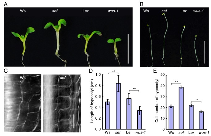
sef is a gain-of-function mutant in which endogenous WUS expression is dramatically elevated; the mutant exhibits clustered ectopic floral buds on the surface of inflorescence stems [19]. Here, we further examined sef, finding that it had elongated hypocotyls compared to wild type Ws. This was the case in both light-grown and dark-grown seedlings (Figure 1A and 1B). Under light conditions, the hypocotyls in sef were about twice as long as those of Ws. By contrast, hypocotyls in the wus-1 loss-of-function mutant were about third shorter than those of wild type Ler (Figure 1D). To investigate the reason underlying the elongated hypocotyl phenotype in sef, we examined the number of epidermal cells in 8-day-old seedlings (Figure 1C and 1E). The sef hypocotyls contained about twice as many cells as those of Ws, whereas wus-1 contained fewer than Ler. The differences of hypocotyl length and cell number in hypocotyl are significant between wild type and mutant (P < 0.05). These results indicate that sef and wus-1 mutants have aberrant hypocotyl lengths due to altered hypocotyl cell production.
Figure 1. Hypocotyl phenotypes of sef and wus-1.
(A) Hypocotyl phenotype of WUS gain-of-function (sef) and loss-of-function (wus-1) mutant seedlings grown in 16-h light/8-h dark. Bar = 1 cm. (B) Hypocotyl length of dark-grown seedlings of sef and wus-1 mutant and their corresponding wild type. Bar = 1 cm. (C) Comparison of the cell number in a same length of hypocotyl in 8-day-old seedlings. Bar = 50 µm. (D) Hypocotyl length of 8-day-old seedlings. Data are means ± SD (n > 15) Student’s t test, **P < 0.01. (E) Hypocotyl cell number of 8-day-old seedlings. Data are means ± SD (n > 15). Student’s t test, **P < 0.01, *P < 0.05.
The cell division rate is increased in sef hypocotyls
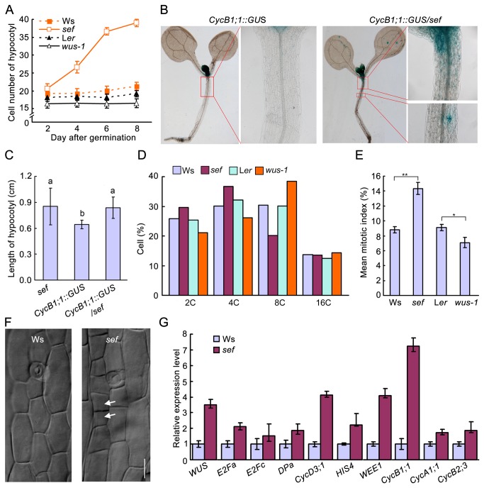
To investigate cell accumulation in the hypocotyl, we monitored cell numbers at different times after germination. Our results showed that cells in the hypocotyl of sef divided faster than those of the wild type at 2, 4 and 6 days after germination (Figure 2A). By contrast, cells in wus-1 and wild-type hypocotyls almost don’t divide during 2- to 8-day after germination. These results suggest that enhanced expression of WUS promotes cell division in the hypocotyl after germination.
Figure 2. Aberrant cell division in hypocotyl of sef.
(A) Cell number of hypocotyl at given days after germination. Data are means ± SD (n > 15). (B) CycB1;1::GUS expression patterns in 8-day-old seedling of CycB1;1::GUS and CycB1;1::GUS/sef. (C) Hypocotyl length of 8-day-old sef, CycB1;1::GUS and CycB1;1::GUS/sef seedlings. Data shown are average values ± SD (n > 15). Different letters represent significant differences according to Student’s t test, *P < 0.05. (D) Cell cycle progression in hypocotyls of Ws, sef, Ler and wus-1 detected by flow cytometry. (E) Mean mitotic index in hypocotyls of Ws, sef, Ler and wus-1. Student’s t test, **P < 0.01, *P < 0.05. (F) Cell division in the hypocotyls of 4-day-old sef seedlings. Bar = 25 µm. (G) Expression levels of cell cycle-related genes in wild type and sef. Data are means ± SD (n = 3).
CycB1;1::GUS is a classic marker used to investigate cell division [20]. We generated Ws and sef plants harboring CycB1;1::GUS. Strong GUS activity was detected in young leaves and the shoot apical meristem of CycB1;1::GUS seedlings (Figure 2B). In CycB1;1::GUS/sef seedlings, GUS activity was additionally observed in the hypocotyls (Figure 2B). The hypocotyls of CycB1;1::GUS/sef seedlings were longer than those of CycB1;1::GUS seedlings, similar to those of sef compared to wild type Ws (Figure 1D and Figure 2C).
To investigate the effect of WUS on cell cycle progression, we measured ploidy levels of hypocotyl cells by flow cytometry. The numbers of 2C and 4C cells were significantly higher in sef than in Ws. In wus-1, there were fewer of both 2C and 4C cells than in wild type Ler. There were fewer 8C cells in sef than in Ws, and more in wus-1 than in Ler. A high fraction of 2C and 4C cells and a low fraction of 8C cells can be indicative of promotion of mitosis [21]. As such, our results suggest that more cells with 2C and 4C in the G2/M phase in sef hypocotyls but less in wus-1 hypocotyls (Figure 2D).
The mitotic index is defined as the ratio of the number of cells in mitosis to the total number of cells and is used as an indicator of the proliferation status in a cell population [21]. The mitotic index in the hypocotyls of sef and wild type was calculated based on the flow cytometric assay. In sef hypocotyls, the mitotic index was significantly higher than in Ws (P < 0.01 by Student’s t test) (Figure 2E). Consistent with this, cell division could be observed in the hypocotyl epidermis of 4-day-old sef seedlings (Figure 2F). This suggests that cytokinesis took place in the hypocotyl of sef.
Expression levels of checkpoint-related genes in cell cycle were analyzed by quantitative RT-PCR (qRT-PCR). The tested genes included: G1-S transition genes E2Fa, E2Fc, DPa and CycD3; 1; S phase gene HIS4; and G2-M transition genes WEE1, CycB1; 1, CycB2;3, and CycA1; 1 [21,22]. Our results demonstrated that in sef, E2Fa and DPa expression increased 2-fold, and that of CycD3; 1 increased by more than 4-fold compared to wild type. We also found that expression of the S phase gene HIS4 was increased about 3-fold in sef compared to wild type. The expression levels of both WEE1 and CycB1; 1 were up regulated more than 4-fold in sef (Figure 2G). These qRT-PCR results showing increased expression of cell cycle-related genes are consistent with cell division taking place in the sef hypocotyl.
We also examined seed and cotyledon size in sef. Compared to wild type, sef seeds and cotyledons were dramatically larger (Figure S1A, S1B and S1C). The size of the palisade cells in sef was similar to that of the Ws (Figure S1E). However, there were more cotyledon cells in sef than in Ws (Figure S1D). These results further confirm that sef has a higher cell division rate than wild type, leading to larger cotyledons as well as longer hypocotyls.
WUS is expressed ectopically in sef hypocotyls
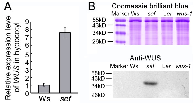
Based on the increased cell division rate in the hypocotyl of sef and the reduced rate in wus-1, we hypothesized that the increased WUS levels might be responsible for the extra cell division in sef. To investigate this, RT-PCR was used to examine the expression of WUS in hypocotyls. Total RNA was isolated from the hypocotyls of 8-day-old Ws and sef seedlings. WUS transcript was detected after 25 cycles in the sef hypocotyl samples, but not in the wild-type samples. At 40 cycles, the amplification of WUS was saturated in sef, but the transcripts was still undetectable in the wild-type hypocotyls (Figure S2A). We also used qRT-PCR to check the transcriptional level of WUS (Figure 3A), and immunoblotting to examine the WUS protein level (Figure 3B) in the sef hypocotyls. Our results showed that both the RNA and protein of WUS were detected in the sef hypocotyls but not in the wild type (Figure 3A and 3B). These data demonstrate that, unlike in wild type, WUS is expressed in sef hypocotyls.
Figure 3. WUS is expressed ectopically in hypocotyls of sef.
(A) Expression of WUS in hypocotyls of Ws, sef, Ler and wus-1 detected by qRT-PCR. Data are means ± SD (n = 3). (B) Immunoblot analysis of WUS protein in hypocotyls of Ws, sef, Ler and wus-1. Upper panel, coomassie brilliant blue (CBB)-stained SDS-PAGE gel. Bottom panel, immunoblotting of WUS protein.
WUS binds the GRP23 promoter directly to activate its expression
WUS directly binds to at least two distinct DNA motifs in the promoters of its target genes, the TAAT motif [23] and TCACGTGA [12]. WUS has been reported to have more than 100 direct targets, including genes involved in development, hormone signaling, and cell division. Based on the presence of these motifs in its promoter, GRP23 is one of the potential direct targets of WUS [12]. Our qRT-PCR analysis revealed that GRP23 expresses not only in flower and root but also in hypocotyl in wild type (Figure S3).
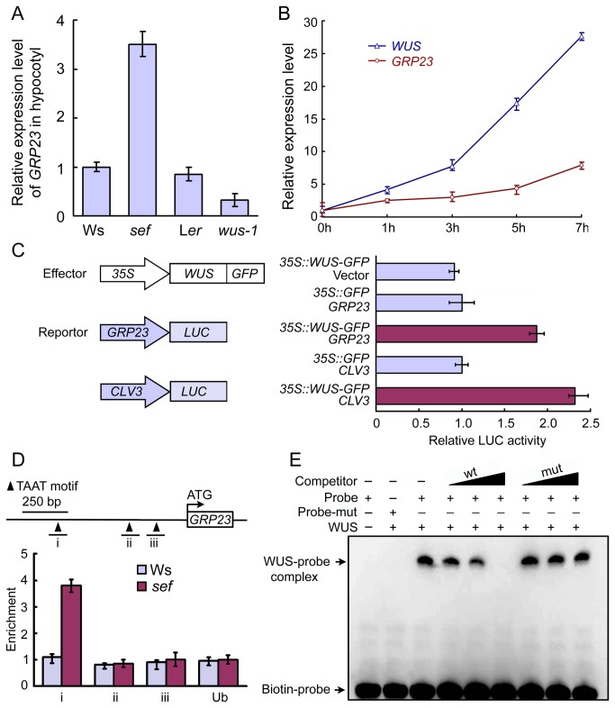
To address the relationship between WUS and GRP23, the expression levels of GRP23 in sef and wus-1 hypocotyls were examined by qRT-PCR. GRP23 transcripts were 3.5-fold more abundant in sef compared to the Ws, whereas in wus-1, GRP23 expression was 0.32-fold that of the Ler (Figure 4A). GRP23 expression was also monitored in pga6-1, an inducible WUS overexpression line [24], after WUS expression was induced with 17-β-estradiol for different lengths of time. The expression of GRP23 increased upon induction of WUS expression in pga6 (Figure 4B).
Figure 4. WUS binds the GRP23 promoter directly to activate its expression.
(A) qRT-PCR analysis of GRP23 expression in Ws, sef, Ler and wus-1. Data are means ± SD (n = 3). (B) qRT-PCR analysis of GRP23 expression in 14-day-old pga6 seedlings after inducing with 17-β-estradiol for 1, 3, 5 and 7 hours. Data are means ± SD (n = 3). (C) Transient expression assay in Arabidopsis protoplasts. The promoters of GRP23 and CLV3 were used to drive the luciferase (LUC) reporter gene. WUS:GFP fusion driven by the 35S promoter was used as effector. LUC activity was assayed after transformation. Data are means ± SD (n = 3). (D) ChIP assay of 7-day-old seedlings to show WUS-binding regions in GRP23 promoter. Regions i, -784 to -660; ii, -311 to -211; iii, -195 to -95. Data are means ± SD (n = 3). (E) EMSA of WUS binding the GRP23 promoter in vitro. The unlabeled double-strands probe (wt) and unlabeled mutant probe (mut) were used for competitive inhibition with 200X, 400X, or 800X molar excess.
To test whether WUS directly binds to the promoter of GRP23, we performed transient expression assays, chromatin immunoprecipitation (ChIP) assays, and electrophoretic mobility shift assays (EMSAs). We performed transient activation assays using protoplasts from Arabidopsis . The LUCIFERASE (LUC) gene driven by the GRP23 promoter (2.0 kb upstream of ATG) was transformed along with various effector constructs into Arabidopsis protoplasts. The promoter of CLV3, a target gene of WUS [25], was used as a positive control. When Arabidopsis protoplasts were co-transfected with the reporter plasmids containing GRP23::LUC or CLV3::LUC and the effector plasmid containing 35S::WUS, the relative LUC activity was increased by 1.9- and 2.4-fold compared to the control (Figure 4C). Thus, our results indicate that WUS serves as an activator for GRP23 transcription in protoplasts.
To further determine whether WUS directly associates with the promoter sequence of GRP23 in vivo, we performed ChIP assay. As shown in Figure 4D, the region “i” of the GRP23 promoter, which included two TAAT motifs from -784 to -660, were enriched with higher abundance in sef compared with Ws.
In EMSA experiments using biotin-labeled fragments with 40 base pairs of GRP23 promoter (-748 to -709), covering two TAAT motifs, a clear WUS-dependent mobility shift was identified (Figure 4E). The unlabeled fragments competitively inhibited this binding. When the TAAT motif was replaced by GGGG, the unlabeled mutated fragments cannot influence the binding of WUS protein (Figure 4E). It is indicated that WUS proteins directly bind to the promoter region of GRP23 in vitro. Taken together, these data suggest that GRP23 expression can be activated by WUS directly in sef hypocotyl.
The expression of GRP23 affects hypocotyl length
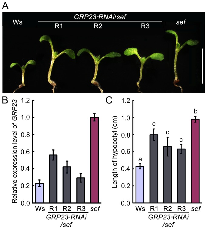
To test the role of GRP23 in hypocotyl growth, we used an RNAi approach to generate three independent GRP23 knockdown transgenic lines in the sef background (GRP23-RNAi/sef). qRT-PCR analysis revealed a reduction of GRP23 transcript to 58%, 44%, and 27% in the three transgenic lines R1, R2, and R3, respectively (Figure 5B and Figure S2B). Hypocotyl length in the transgenic lines was intermediate between those of Ws and sef (Figure 5A and 5C). These results demonstrate that knockdown of GRP23 can partially attenuate the elongated hypocotyl phenotype of sef.
Figure 5. Knockdown of GRP23 partially rescues the sef phenotype.
(A) Hypocotyl phenotype of Ws, sef and sef GRP23-RNAi seedlings after germination for 8 days. Bar = 1 cm. R1, R2, and R3 represent the RNAi lines 1, 2, and 3 respectively. (B) Expression of GRP23 in sef and GRP23-RNAi/sef seedlings detected by qRT-PCR. Data are means ± SD (n = 3). (C) Hypocotyl length of 8-day-old seedlings of Ws, sef and GRP23-RNAi/sef. Data are means ± SD (n > 15). Different letters a, b, and c represent significantly differences among the lines (*P < 0.05) by Student’s t test.
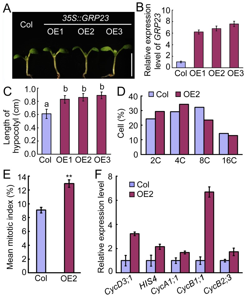
To confirm the function of GRP23 in hypocotyl cell division, three independent 35S::GRP23 transgenic lines, OE1, OE2, and OE3 were obtained. GRP23 transcript was markedly increased in all three lines compared with wild type (Figure 6B). In addition, hypocotyl length in the three 35S::GRP23 lines was significantly increased compared to wild type (P < 0.05) (Figure 6A and 6C). These results indicate that GRP23 overexpression can mimic the elongated hypocotyl phenotype of sef.
Figure 6. Phenotype of 35S::GRP23 transgenic plants.
(A) Hypocotyl phenotype of Col and 35S::GRP23 transgenic lines (OE1, OE2, and OE3) after germination for 8 days. Bars = 1 cm.(B) GRP23 expression in Col and 35S::GRP23 seedlings detected by qRT-PCR. Data are means ± SD (n = 3). (C) Hypocotyl length of Col and 35S::GRP23 seedlings after germination for 8 days. Data are means ± SD (n > 15). Different letters a and b represent significantly differences among the lines (*P < 0.05) by Student’s t test. (D) Cell cycle progression in hypocotyls of Col and 35S::GRP23 line 2 (OE2) analyzed by flow cytometry. (E) Mean mitotic index in hypocotyl of Col and 35S::GRP23 line 2. Student’s t test, **P < 0.01. (F) Expression levels of cell cycle-related genes in Col and 35S::GRP23 line 2. Data are means ± SD (n = 3).
We also performed flow cytometry assays to examine the cell cycle in hypocotyls of OE2. The hypocotyls of 35S::GRP23 plants possessed more cells with 2C or 4C in the G2/M phase and had a higher mitotic index than those of the wild type (Figure 6D and 6E). The expression levels of cell cycle checkpoint-related genes were elevated in 35S::GRP23 hypocotyls compared to those of wild type (Figure 6F). These data suggest that GRP23 promotes cell division in the hypocotyl through controlling the G2/M transition.
Discussion
Aberrantly long hypocotyls in sef are caused by ectopic expression of WUS
WUS specifies stem cell identity in the cells overlying of the central zone, and is both necessary and sufficient for stem cell maintenance [2,4]. Moreover, WUS is connected with CLV3 through a regulatory loop for maintaining a constant number of stem cells [2,26,27]. Previous studies have mainly concentrated on the mechanism through which WUS maintains the number of stem cells in the shoot and floral meristems. Here we report the effect of ectopic WUS on cell division in hypocotyl.
Cell number in the hypocotyl is constant, with approximately 20 cells in Arabidopsis [14,16]. Although a considerable number of mutants with altered hypocotyl length have been studied, these reports all focused on cell elongation [15,28]. For example, Arabidopsis ENHANCED PHOTOMORPHOGENIC 1 (EPP1) encodes an ATP-dependent chromatin remodeling factor. EPP1 interacts with HY5 to regulate cell elongation in the hypocotyl [29]. MICROTUBULE-DESTABILIZING PROTEIN 25 (MDP25) modulates hypocotyl length by affecting cell elongation [30]. By contrast, our results revealed that the long and short hypocotyls in sef and wus-1, respectively, were caused not by differential cell elongation, but by the presence of a different number of cells (Figure 1). In addition, more cells were also found in seeds and cotyledons of sef (Figure S1). WUS is normally expressed in cells of the organizing center and in overlying cells of the central zone [2,4]. We speculated that WUS might be ectopically expressed in sef hypocotyls based on the previously established ectopic WUS expression in the inflorescence stem of sef [19]. Indeed, we found evidences of WUS expression at both the transcript and protein level in hypocotyls of the sef mutant (Figure 3). It is likely that the ectopic WUS expression in sef is due to insertion of 35S enhancers in the WUS promoter [19]. Other previous reports also showed a similar phenomenon, in wild-type Arabidopsis , no transcripts of the HOMEODOMAIN GLABROUS 11 (HDG11) gene can be detected in roots, leaves, or stems, but 35S enhancers resulted in the overexprssion of HDG11 in a constitutive fashion [31]. Together, these results indicate that WUS is involved in controlling hypocotyl length in sef by altering cell number. The increase of cell number in sef hypocotyls resulted from the activation of GRP23 by ectopic WUS.
WUS directly represses the transcription of ARR5, ARR6 and ARR7, which act as negative regulators of cytokinin signaling [6]. The expression of ARR5, ARR6 and ARR7 was inhibited in sef hypocotyls (Figure S4). Based on these results, it is likely that ectopic expression of WUS in sef hypocotyls results in an enhanced cytokinin signal to activate cell division.
WUS regulates GRP23 to mediate cell division in the hypocotyl
Post embryonic growth of Arabidopsis hypocotyl, cell division in the hypocotyl occurs only in the epidermis during the formation of stomata. The elongation of hypocotyls does not involve cell division in the cortex or epidermis [14,16,32]. However, our results showed that the increase in cell number of sef hypocotyls occurred mainly during postembryonic development (Figure 2A). Reporter gene expression levels and flow cytometry assay results also supported the idea that cell division occurred in sef hypocotyl due to WUS ectopic expression (Figures 2, 3). Ectopic expression of WUS driven by the 35S promoter occasionally causes activation of the CycB1;1::GUS reporter gene along the vasculature of leaves [33]. These results indicate that WUS can promote cell division in tissues outside of the organizing center of stem cells in Arabidopsis .
The pathway through which WUS activates cell division is unknown. However ChIP-chip results revealed that GRP23 is one of the 159 direct WUS target genes and can be induced by WUS in Arabidopsis apices [12]. Moreover, the histochemical assay of GRP23::GUS and in situ hybridization showed that GRP23 expresses in the embryo, ovules, primordium of leaf and lateral root, and apical meristems of root and shoot [13]. The expression patterns of GRP23 and WUS overlap in embryo and shoot meristem [2,3,13]. Based on these results it can be speculated that GRP23 is a direct target of WUS in the wild-type meristem. Our ChIP, EMSA and LUC activity results showed that WUS directly binds the GRP23 promoter to activate reporter gene expression (Figure 4). Our results are consistent with the previous findings of Busch et al. [12]. Together, these results indicate that WUS directly targets GRP23 to activate its expression both in wild-type shoot apical meristem and in sef hypocotyl.
Reduced expression of GRP23 rescued the elongated hypocotyl phenotype of sef, whereas GRP23 overexpression resulted in a higher mitotic index and increased expression of cell division related genes, mimicking sef (Figure 5 and Figure 6). These results support the idea that WUS promotes cell division via GRP23, which encodes a PENTATRICOPEPTIDE REPEAT (PPR) protein. The grp23 mutant displays an aberrant cell division pattern [13]. Mutants of another PPR protein gene, PPR2263, exhibit growth defects and reduced size resulting from altered cell division [34]. These reports are consistent with our observation that the PPR protein GRP23 promotes cell division.
In conclusion, our data suggest that ectopic expression of WUS in hypocotyl regulates cell division via promoting GRP23 expression. GRP23 is a direct target gene of transcription factor WUS that mediates it effects on cell division in hypocotyls.
Materials and Methods
Plant materials and growth conditions
The gain-of-function mutant sef (ecotype Ws-2) was identified via activation tagged genetic screening as described previously [19]. wus-1 (ecotype Ler) was obtained from the Arabidopsis Biological Resource Center at Ohio State University (Columbus, USA). The pga6-1 mutant and 35S::GRP23 transgenic plants were kindly provided by Prof. Jianru Zuo [24] and Prof. Weicai Yang [13] (Institute of Genetics and Developmental Biology, CAS, Beijing, China). Seeds were surface-sterilized with 10% bleach plus 0.01% Triton X-100 for 15 min, and then washed four times with sterile water. The surface-sterilized seeds were stratified at 4 °C for 2 days and transferred to medium or soil for further growth (16-h light/8-h dark, 22°C). pga6-1 was treated with 10 µM 17-β-estradiol for different amounts of time as described by Zuo et al. [24].
Measurement of length and cell number in hypocotyls
For phenotype analysis, seedlings were grown on 0.8% phytoagar plates containing half-strength Murashige-Skoog nutrients and 1% sucrose. Image J1.34 (http://rsb.info.nih.gov/ij/download.html) was used to measure hypocotyl length, seed size, and cotyledon size after photographing. Hypocotyl length was measured from the base of the cotyledon to the junction of the hypocotyl and the primary root.
To count the cell number, 2- to 8-day-old seedlings, were mounted with a clearing solution [35]. After 15–60 min, samples were examined under microscope (Lecia DM2500, Germany). The cell numbers in cotyledons and hypocotyls were counted. Quantitative data were subjected to two-tailed independent Student’s t tests using SPSS 18.0 software (http://www.spss.com). Significance levels of P < 0.05 and P < 0.01 are indicated by single and double asterisks, respectively.
Flow cytometry analysis
For flow cytometry analysis, seedlings were plated onto half-strength Murashige-Skoog media. After 8 days in the greenhouse (16-h light/8-h dark, 22°C), hypocotyls were collected for flow cytometry analysis as previously described by Galbraith et al. [36]. The nuclei were analyzed with a ploidy analyzer FACS Caliber (BD Corporation). At least three biological replicates were used for each sample.
RT-PCR and quantitative RT-PCR (qRT-PCR)
Total RNA was isolated from 8-day-old Arabidopsis hypocotyls or seedlings using TRIzol reagent (Invitrogen). The DNase-treated RNA was reverse-transcribed using M-MLV reverse transcriptase (Promega). cDNAs were synthesized from 2.0 µg total RNA using Superscript reverse transcriptase. RT-PCR was performed with gene-specific primers (Table 1) and runs 18-40 cycles depending on the linear range of products for each gene. RT-PCR reactions were repeated five times.
Table 1. Primer sequences.
| qRT-PCR | |
|---|---|
| WUS-F | 5'-GCTAATTCCGTCAACGTTAAAC-3’ |
| WUS-R | 5'-TTTAAATTCCCGTTATTGAAGC-3’ |
| WEE1-F | 5’-TTGGACAAAAGCTTACCAGTAGAAG-3’ |
| WEE1-R | 5’-AGAGAAGATATCGACTTTATCAAGG-3’ |
| HIS4-F | 5’-TTAGGCAAAGGAGGAGCAAA-3’ |
| HIS4-R | 5’-CTCCTCGCATGCTCAGTGTA-3’ |
| CycD3;1-F | 5’-GCAAGTTGATCCCTTTGACC-3’ |
| CycD3;1-R | 5’-CAGCTTGGACTGTTCAACGA-3’ |
| CycB1;1-F | 5’-CTCAAAATCCCACGCTTCTTGTGG-3’ |
| CycB1;1-R | 5’-CACGTCTACTACCTTTGGTTTCCC-3’ |
| CycA1;1-F | 5’-GGCTAAGAAGCGACCTGATG-3’ |
| CycA1;1-R | 5’-TACAAGCCACACCAAGCAAC-3’ |
| CycB2;3-F | 5’-TAAACCACCTGTGCATCGAC-3’ |
| CycB2;3-R | 5’-ATCTCCTCCAGCATTGCTTC-3’ |
| E2Fa-F | 5’-ACGCTGGTTCTCCTATCACAC-3’ |
| E2Fa-R | 5’-GGCTTGTTTAATTAGATTGACGAA-3’ |
| E2Fc-F | 5’-GGAAGGGTGCTGACAATCTT-3’ |
| E2Fc-R | 5’-CATCCAACCTGCTTTCCTCA-3’ |
| DPa-F | 5’-GATGATTCTGAAATTGGATCAGAG-3’ |
| DPa-R | 5’-TTGGCTTCCAACTTCTGACA-3’ |
| GRP23-F | 5’-TGCTCCATCCTCAGTTACTT-3’ |
| GRP23-R | 5’-AATAAACTCGCAGCATCTCC-3’ |
| ACTIN-F | 5’-GCTCCTCTTAACCCAAAGGC-3’ |
| ACTIN-R | 5’-CACACCATCACCAGAATCCAGC-3’ |
| ARR5-F | 5'-TTTGCGTCCCGAGATGTTAG-3’ |
| ARR5-R | 5'-CCATACTATCATCAACAGCAAGAAC-3’ |
| ARR6-F | 5’-TTGCCTCGTATTGATAGATGTC-3’ |
| ARR6-R | 5’-CGAGTGAACAGGGTAGACATT-3’ |
| ARR7-F | 5’-AATGCCAGGACTTTCAGGAT-3’ |
| ARR7-R | 5’-ATTCCTCTGCTCCTTCTTTG-3’ |
| RT-PCR | |
| WUS-pBI221-F | 5'-CCGCTCGAGATGGAGCCGCCACAGCATCAGCATC-3’ |
| WUS-pBI221-R | 5'-GGGGTACCCTAGTTCAGACGTAGCTCAAGAGAA-3’ |
| GRP23-LUC-F | 5’-CGGGATCCTATCCAGCTAATCCCATCTGCTCTT-3’ |
| GRP23-LUC-R | 5’-CCCAAGCTTGGTGGAGGGAAAATGATTTAGGGTT-3’ |
| CLV3-LUC-F | 5’-CGGGATCCGCAACCTTCGATAGAAATAGTGAC-3’ |
| CLV3-LUC-R | 5’-CCCAAGCTTAAGACACAAGTATATCTCCAAAGC-3’ |
| GRP23 RNAi-F | 5’-GGCGCGCCGGATCCCGGAGATGCTGCGAGTT-3’ |
| GRP23 RNAi-R | 5’-CATGCCATGGTCTAGACTTACTTCCGACCTTCTT-3’ |
| ACTIN-F | 5’-TTTGCGACAATGGAACTG-3’ |
| ACTIN-R | 5’-AAGAGCAATGTAGCAAAG-3’’ |
| EMSA | |
| GRP23p-Probe-F | 5'-CATATATCTTTAATACTGTTAATGATCTTTCTTCAAAAAC-3’ |
| GRP23p-Probe-R | 5'-GTTTTTGAAGAAAGATCATTAACAGTATTAAAGATATATG-3’ |
| ChIP | |
| GRP23p i-F | 5'-TCACGTTATATGAGCATCTTTT-3’ |
| GRP23p i-R | 5'-TTGAAACTGA AACTTTATACGAAA-3’ |
| GRP23p ii-F | 5'-ACCAGCTATGGATTATTTGAGA-3’ |
| GRP23p ii-R | 5'-AAGACAAGTAAAGAAAGGTTGG-3’ |
| GRP23p iii-F | 5'-CGTATTACCAAACAGCCCTC-3’ |
| GRP23p iii-R | 5'-CCTTGGATGTGAAGAAATGG-3’ |
| UBQ-F | 5'-CAGGATAAGGAGGGCATT-3’ |
| UBQ-R | 5'-TTTCCCAGTCAACGTCTT-3’ |
qRT-PCR was performed on an Applied Biosystems 7500 real time PCR System using SYBR Premix Ex Taq™ (TaKaRa). The following thermal cycle was used: 95°C for 3 min, then 40 cycles of 95°C for 30 s, 60°C for 30 s, and 72°C for 1 min. The Actin1 gene (accession no. X16280) was used as the internal control. The relative expression levels were analyzed using a relative quantitation method (∆∆CT) for every PCR. The primers used for qRT-PCR are listed in Table 1.
Immunoblotting
Total protein samples were extracted from 8-day-old Arabidopsis hypocotyls as described previously [37]. Proteinase inhibitors were added and proteins were separated on 12% SDS-PAGE gels and then transferred to a polyvinylidene fluoride (BioTraceTM, USA) membrane. Membranes were blocked for 1 h with 5% BSA in TBS-Tween buffer (Tris-HCl 20 mM, NaCl 150 mM, and Tween 0.05%, pH 8.0). Immunoprobing of WUS was conducted with the rabbit anti-WUS (A gift from Huiqin Ma) (1:3,000) polyclonal antibody in TBS. An anti-rabbit IgG (1:10,000) conjugated with alkaline phosphatase was used as the secondary antibody with an ECL protein gel blot detection system (Amersham, Sweden).
LUC activity assay
Protoplast isolation and transient expression assays were performed as described by Lin et al. [38]. GRP23 and CLV3 promoters (2kb) were amplified from genomic DNA and inserted into the reporter plasmid to drive the expression of LUC. To produce the effector plasmid, the full-length WUS CDS was inserted into the pBI221 plasmid and driven by CaMV 35S promoter. The primers used for amplification were listed in Table 1. For transient expression assays, the reporter plasmids pYY96-GRP23::LUC or pYY96-CLV3::LUC and effector constructs pBI221-WUS were cotransformed into protoplasts. The reporter gene GUS driven by 35S promoter was used as an internal control to normalize LUC expression. GUS fluorescence was detected with a UV fluorescence optical kit using a GLOMAX 20/20 LUMINOMETER (Promega). LUC activity was measured using LUC assay substrate with a luminescence kit (Promega). The relative reporter gene expression levels were expressed as the LUC/GUS ratios.
Chromatin immunoprecipitation assays
Chromatin immunoprecipitation (ChIP) was performed as described with 7-day-old seedlings [39]. The rabbit anti-WUS polyclonal antibody was used for immunoprecipitation. ChIP products were analyzed by qRT-PCR, and the enriched relative abundance was expressed as the ratio of sef to Ws. Data are means ± SD of three independent experiments.
EMSA
EMSA was performed essentially as described [40]. Briefly, the coding sequence of WUS was cloned into the expression vector pGEX-4T-1. The recombinant pGEX-4T-1-WUS was transformed into Escherichia coli BL21. Cells were grown at 37°C and induced by 1 mM isopropyl β-D-1-thiogalactopyranoside for 5 h and purified by glutathione affinity chromatography as described in the Bulk and RediPack GST purification kit (Pharmacia). EMSAs were performed using the biotin-labeled probes and the Lightshift Chemiluminescent EMSA kit (Pierce) according to the manufacturer’s instructions. Wild-type and mutated oligonucleotides were synthesized as single-stranded DNA. The wild-type oligonucleotide sequence with 40 bases corresponds to the -748 to -709 regions in the GRP23 promoter. In the mutated oligonucleotide, two TAAT motifs (-738 to -735 and -729 to -726) were replaced by GGGG. Single-strand oligonucleotides were labeled with biotin at 3'-end, and then equal amounts of labeled complementary oligonucleotides were mixed, boiled for 2 min, and then slowly cooled down to 25°C for annealing. The labeled double-strand fragments were detected according to the instructions provided with the EMSA kit (Pierce). For competition experiments, different amounts of unlabeled wild-type and mutated double-strand fragments were added to the binding reaction.
Supporting Information
Seed and cotyledon phenotypes of sef.
(DOCX)
Expression levels of WUS and GRP23 detected by RT-PCR.
(DOCX)
Expression levels of GRP23 in various tissues of Arabidopsis .
(DOCX)
Expression levels of A-type ARRs genes in hypocotyl.
(DOCX)
Acknowledgments
We are grateful to Professor NamHai Chua, Jianru Zuo and Weicai Yang for sharing pga6-1 mutant seeds and 35S::GRP23 transgenic seeds. We also thank Huiqin Ma (Chinese Agriculture University) for the WUS antiserum. We would like to thank Dr. JH Snyder, Dr. N Hofmann, and Mr. GT Sniffen for examining the English usage in the manuscript, as well as Yong Hu (Capital Normal University, Beijing) and Suhua Yang (Key Lab of Plant Molecular Physiology, Institute of Botany, CAS) for their help in flow cytometric analysis.
Funding Statement
This work was supported by the National Science Foundation of China for Innovative Research Groups (31121065). The funders had no role in study design, data collection and analysis, decision to publish, or preparation of the manuscript.
References
- 1. Gutierrez C (2005) Coupling cell proliferation and development in plants. Nat Cell Biol 7: 535-541. doi:10.1038/ncb0605-535. PubMed: 15928697. [DOI] [PubMed] [Google Scholar]
- 2. Mayer KF, Schoof H, Haecker A, Lenhard M, Jürgens G et al. (1998) Role of WUSCHEL in regulating stem cell fate in the Arabidopsis shoot meristem. Cell 95: 805-815. doi:10.1016/S0092-8674(00)81703-1. PubMed: 9865698. [DOI] [PubMed] [Google Scholar]
- 3. Schoof H, Lenhard M, Haecker A, Mayer KF, Jürgens G et al. (2000) The stem cell population of Arabidopsis shoot meristems in maintained by a regulatory loop between the CLAVATA and WUSCHEL genes. Cell 100: 635-644. doi:10.1016/S0092-8674(00)80700-X. PubMed: 10761929. [DOI] [PubMed] [Google Scholar]
- 4. Laux T, Mayer KF, Berger J, Jürgens G (1996) The WUSCHEL gene is required for shoot and floral meristem integrity in Arabidopsis . Development 122: 87-96. PubMed: 8565856. [DOI] [PubMed] [Google Scholar]
- 5. Chickarmane VS, Gordon SP, Tarr PT, Heisler MG, Meyerowitz EM (2012) Cytokinin signaling as a positional cue for patterning the apical-basal axis of the growing Arabidopsis shoot meristem. Proc Natl Acad Sci U S A 109: 4002-4007. doi:10.1073/pnas.1200636109. PubMed: 22345559. [DOI] [PMC free article] [PubMed] [Google Scholar]
- 6. Leibfried A, To JPC, Busch W, Stehling S, Kehle A et al. (2005) WUSCHEL controls meristem function by direct regulation of cytokinin-inducible response regulators. Nature 438: 1172-1175. doi:10.1038/nature04270. PubMed: 16372013. [DOI] [PubMed] [Google Scholar]
- 7. Buechel S, Leibfried A, To JP, Zhao Z, Andersen SU et al. (2009) Role of A-type Arabidopsis RESPONSE REGULATORS in meristem maintenance and regeneration. Eur J Cell Biol 89: 279-284. PubMed: 20018401. [DOI] [PubMed] [Google Scholar]
- 8. Cheng ZJ, Wang L, Sun W, Zhang Y, Zhou C et al. (2013) Pattern of auxin and cytokinin responses for shoot meristem induction results from the regulation of cytokinin biosynthesis by AUXIN RESPONSE FACTOR3. Plant Physiol 161: 240-251. doi:10.1104/pp.112.203166. PubMed: 23124326. [DOI] [PMC free article] [PubMed] [Google Scholar]
- 9. Gordon SP, Chickarmane VS, Ohno C, Meyerowitz EM (2009) Multiple feedback loops through cytokinin signaling control stem cell number within the Arabidopsis shoot meristem. Proc Natl Acad Sci U S A 106: 16529-16534. doi:10.1073/pnas.0908122106. PubMed: 19717465. [DOI] [PMC free article] [PubMed] [Google Scholar]
- 10. Sablowski R (2009) Cytokinin and WUSCHEL tie the knot around plant stem cells. Proc Natl Acad Sci U S A 106: 16016-16017. doi:10.1073/pnas.0909300106. PubMed: 19805255. [DOI] [PMC free article] [PubMed] [Google Scholar]
- 11. Müller B, Sheen J (2008) Cytokinin and auxin interaction in root stem-cell specification during early embryogenesis. Nature 453: 1094-1097. doi:10.1038/nature06943. PubMed: 18463635. [DOI] [PMC free article] [PubMed] [Google Scholar]
- 12. Busch W, Miotk A, Ariel FD, Zhao Z, Forner J et al. (2010) Transcriptional control of a plant stem cell niche. Dev Cell 18: 849-861. PubMed: 20493817. [DOI] [PubMed] [Google Scholar]
- 13. Ding YH, Liu NY, Tang ZS, Liu J, Yang WC (2006) Arabidopsis GLUTAMINE-RICH PROTEIN23 is essential for early embryogenesis and encodes a novel nuclear PPR motif protein that interacts with RNA polymerase II subunit III. Plant Cell 18: 815-830. doi:10.1105/tpc.105.039495. PubMed: 16489121. [DOI] [PMC free article] [PubMed] [Google Scholar]
- 14. Gendreau E, Traas J, Desnos T, Grandjean O, Caboche M et al. (1997) Cellular basis of hypocotyl growth in Arabidopsis thaliana . Plant Physiol 114: 295-305. doi:10.1104/pp.114.1.295. PubMed: 9159952. [DOI] [PMC free article] [PubMed] [Google Scholar]
- 15. Clouse SD (1996) Molecular genetic studies confirm the role of brassinosteroids in plant growth and development. Plant J 10: 1-8. doi:10.1046/j.1365-313X.1996.10010001.x. PubMed: 8758975. [DOI] [PubMed] [Google Scholar]
- 16. Collett CE, Harberd NP, Leyser O (2000) Hormonal interactions in the control of Arabidopsis hypocotyl elongation. Plant Physiol 124: 553-561. doi:10.1104/pp.124.2.553. PubMed: 11027706. [DOI] [PMC free article] [PubMed] [Google Scholar]
- 17. Cowling RJ, Harberd NP (1999) Gibberellins control Arabidopsis hypocotyl growth via regulation of cellular elongation. J Exp Bot 50: 1351-1357. doi:10.1093/jexbot/50.337.1351. [Google Scholar]
- 18. Quail PH, Boylan MT, Parks BM, Short TW, Xu Y et al. (1995) Phytochromes-photosensory perception and signal-transduction. Science 268: 675-680. doi:10.1126/science.7732376. PubMed: 7732376. [DOI] [PubMed] [Google Scholar]
- 19. Xu YY, Wang XM, Li J, Li JH, Wu JS et al. (2005) Activation of the WUS gene induces ectopic initiation of floral meristems on mature stem surface in Arabidopsis thaliana . Plant Mol Biol 57: 773-784. doi:10.1007/s11103-005-0952-9. PubMed: 15952065. [DOI] [PubMed] [Google Scholar]
- 20. Colón-Carmona A, You R, Haimovitch-Gal T, Doerner P (1999) Spatio-temporal analysis of mitotic activity with a labile cyclin-GUS fusion protein. Plant J 20: 503-508. doi:10.1046/j.1365-313x.1999.00620.x. PubMed: 10607302. [DOI] [PubMed] [Google Scholar]
- 21. Inzé D, De Veylder L (2006) Cell cycle regulation in plant development. Annu Rev Genet 40: 77-105. doi:10.1146/annurev.genet.40.110405.090431. PubMed: 17094738. [DOI] [PubMed] [Google Scholar]
- 22. Dewitte W, Murray JA (2003) The plant cell cycle. Annu Rev Plant Biol 54: 235-264. doi:10.1146/annurev.arplant.54.031902.134836. PubMed: 14502991. [DOI] [PubMed] [Google Scholar]
- 23. Lohmann JU, Hong RL, Hobe M, Busch MA, Parcy F et al. (2001) A molecular link between stem cell regulation and floral patterning in Arabidopsis . Cell 105: 793-803. doi:10.1016/S0092-8674(01)00384-1. PubMed: 11440721. [DOI] [PubMed] [Google Scholar]
- 24. Zuo JR, Niu QW, Frugis G, Chua NH (2002) The WUSCHEL gene promotes vegetative-to-embryonic transition in Arabidopsis . Plant J 30: 349-359. doi:10.1046/j.1365-313X.2002.01289.x. PubMed: 12000682. [DOI] [PubMed] [Google Scholar]
- 25. Yadav RK, Perales M, Gruel J, Girke T, Jönsson H et al. (2011) WUSCHEL protein movement mediates stem cell homeostasis in the Arabidopsis shoot apex. Genes Dev 25: 2025-2030. doi:10.1101/gad.17258511. PubMed: 21979915. [DOI] [PMC free article] [PubMed] [Google Scholar]
- 26. Brand U, Fletcher JC, Hobe M, Meyerowitz EM, Simon R (2000) Dependence of stem cell fate in Arabidopsis an a feedback loop regulated by CLV3 activity. Science 289: 617-619. doi:10.1126/science.289.5479.617. PubMed: 10915624. [DOI] [PubMed] [Google Scholar]
- 27. Fletcher LC, Brand U, Running MP, Simon R, Meyerowitz EM (1999) Signaling of cell fate decisions by CLAVATA3 in Arabidopsis shoot meristems. Science 283: 1911-1914. doi:10.1126/science.283.5409.1911. PubMed: 10082464. [DOI] [PubMed] [Google Scholar]
- 28. McNellis TW, Deng XW (1995) Light control of seedling morphogenetic pattern. Plant Cell 7: 1749-1761. doi:10.1105/tpc.7.11.1749. PubMed: 8535132. [DOI] [PMC free article] [PubMed] [Google Scholar]
- 29. Jing Y, Zhang D, Wang X, Tang W, Wang W et al. (2013) Arabidopsis Chromatin Remodeling Factor PICKLE Interacts with Transcription Factor HY5 to Regulate Hypocotyl Cell Elongation. Plant Cell 25: 242-256. doi:10.1105/tpc.112.105742. PubMed: 23314848. [DOI] [PMC free article] [PubMed] [Google Scholar]
- 30. Li J, Wang X, Qin T, Zhang Y, Liu X et al. (2011) MDP25, a novel calcium regulatory protein, mediates hypocotyl cell elongation by destabilizing cortical microtubules in Arabidopsis . Plant Cell 23: 4411-4427. doi:10.1105/tpc.111.092684. PubMed: 22209764. [DOI] [PMC free article] [PubMed] [Google Scholar]
- 31. Yu H, Chen X, Hong Y-Y, Wang Y, Xu P et al. (2008) Activated expression of an Arabidopsis HD-START protein confers drought tolerance with improved root system and reduced stomatal density. Plant Cell 20: 1134-1151. doi:10.1105/tpc.108.058263. PubMed: 18451323. [DOI] [PMC free article] [PubMed] [Google Scholar]
- 32. Saibo NJM, Vriezen WH, Beemster GTS, Van der Straeten D (2003) Growth and stomata development of Arabidopsis hypocotyls are controlled by gibberellins and modulated by ethylene and auxins. Plant J 33: 989-1000. doi:10.1046/j.1365-313X.2003.01684.x. PubMed: 12631324. [DOI] [PubMed] [Google Scholar]
- 33. Lenhard M, Jürgens G, Laux T (2002) The WUSCHEL and SHOOTMERISTEMLESS genes fulfil complementary roles in Arabidopsis shoot meristem regulation. Development 129: 3195-3206. PubMed: 12070094. [DOI] [PubMed] [Google Scholar]
- 34. Sosso D, Mbelo S, Vernoud V, Gendrot G, Dedieu A et al. (2012) PPR2263, a DYW-subgroup pentatricopeptide repeat protein, is required for mitochondrial nad5 and cob transcript editing, mitochondrion biogenesis, and maize growth. Plant Cell 24: 676-691. doi:10.1105/tpc.111.091074. PubMed: 22319053. [DOI] [PMC free article] [PubMed] [Google Scholar]
- 35. Sabatini S, Beis D, Wolkenfelt H, Murfett J, Guilfoyle T et al. (1999) An auxin-dependent distal organizer of pattern and polarity in the Arabidopsis root. Cell 99: 463-472. doi:10.1016/S0092-8674(00)81535-4. PubMed: 10589675. [DOI] [PubMed] [Google Scholar]
- 36. Galbraith DW, Harkins KR, Maddox JM, Ayres NM, Sharma DP et al. (1983) Rapid flow cytometric analysis of the cell-cycle intact plant-tissues. Science 220: 1049-1051. doi:10.1126/science.220.4601.1049. PubMed: 17754551. [DOI] [PubMed] [Google Scholar]
- 37. Yanagisawa S, Yoo SD, Sheen J (2003) Differential regulation of EIN3 stability by glucose and ethylene signalling in plants. Nature 425: 521-525. doi:10.1038/nature01984. PubMed: 14523448. [DOI] [PubMed] [Google Scholar]
- 38. Lin R, Ding L, Casola C, Ripoll DR, Feschotte C et al. (2007) Transposase-derived transcription factors regulate light signaling in Arabidopsis . Science 318: 1302-1305. doi:10.1126/science.1146281. PubMed: 18033885. [DOI] [PMC free article] [PubMed] [Google Scholar]
- 39. Bowler C, Benvenuto G, Laflamme P, Molino D, Probst AV, Tariq M et al. (2004) Chromatin techniques for plant cells. Plant J 39: 776-789. doi:10.1111/j.1365-313X.2004.02169.x. PubMed: 15315638. [DOI] [PubMed] [Google Scholar]
- 40. Ma QB, Dai XY, Xu YY, Guo J, Liu Y et al. (2009) Enhanced tolerance to chilling stress in OsMYB3R-2 transgenic rice is mediated by alteration in cell cycle and ectopic expression of stress genes. Plant Physiol 150: 244-256. doi:10.1104/pp.108.133454. PubMed: 19279197. [DOI] [PMC free article] [PubMed] [Google Scholar]
Associated Data
This section collects any data citations, data availability statements, or supplementary materials included in this article.
Supplementary Materials
Seed and cotyledon phenotypes of sef.
(DOCX)
Expression levels of WUS and GRP23 detected by RT-PCR.
(DOCX)
Expression levels of GRP23 in various tissues of Arabidopsis .
(DOCX)
Expression levels of A-type ARRs genes in hypocotyl.
(DOCX)


