From: General properties of armed effector T cells

NCBI Bookshelf. A service of the National Library of Medicine, National Institutes of Health.

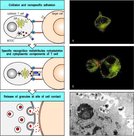
The example illustrated here is a CD8 cytotoxic T cell. Cytotoxic CD8 cells contain specialized lysosomes called lytic granules, which contain cytotoxic proteins. Initial binding to a target cell through adhesion molecules does not have any effect on the location of the lytic granules. Binding of the T-cell receptor causes the T cell to become polarized: reorganization within the cortical actin cytoskeleton at the site of contact has the effect of aligning the microtubule-organizing center (MTOC), which in turn aligns the secretory apparatus, including the Golgi apparatus (GA), towards the target cell. Proteins stored in lytic granules derived from the Golgi are then directed specifically onto the target cell. The photomicrograph in panel a shows an unbound, isolated cytotoxic T cell. The microtubule cytoskeleton is stained in green and the lytic granules in red. Note how the lytic granules are dispersed throughout the T cell. Panel b depicts a cytotoxic T cell bound to a (larger) target cell. The lytic granules are now clustered at the site of cell-cell contact in the bound T cell. The electron micrograph in panel c shows the release of granules from a cytotoxic T cell. Panels a and b courtesy of G. Griffiths. Panel c courtesy of E.R. Podack.
From: General properties of armed effector T cells

NCBI Bookshelf. A service of the National Library of Medicine, National Institutes of Health.