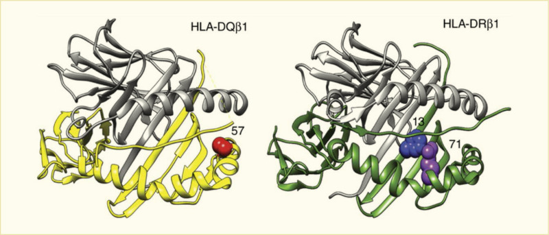
Two panel illustration. Left – H L A D Q molecule structure with amino acid at position 57. Right – H L A D R molecule structure with amino acids at positions 13 and 71.

FIGURE 5.

Structural Models of HLA-DQ and HLA-DR Molecules.

HLA-DQβ1 position 57, HLA-DRβ1 position 13, and HLA-DRβ1 position 71 are each located in the respective molecule’s peptide-binding groove. HLA-DRβ1 positions 13 and 71 line the P4 pocket of the HLA-DR molecule. HLA, human leukocyte antigen.

SOURCE: Reference 33, copyright © 2015 Nature Publishing Group, reprinted with permission.

From: Genetics of Type 1 Diabetes

Cover of Diabetes in America
Diabetes in America [Internet].
Lawrence JM, Casagrande SS, Herman WH, et al., editors.
Copyright Notice

Diabetes in America is in the public domain of the United States. You may use the work without restriction in the United States.

NCBI Bookshelf. A service of the National Library of Medicine, National Institutes of Health.