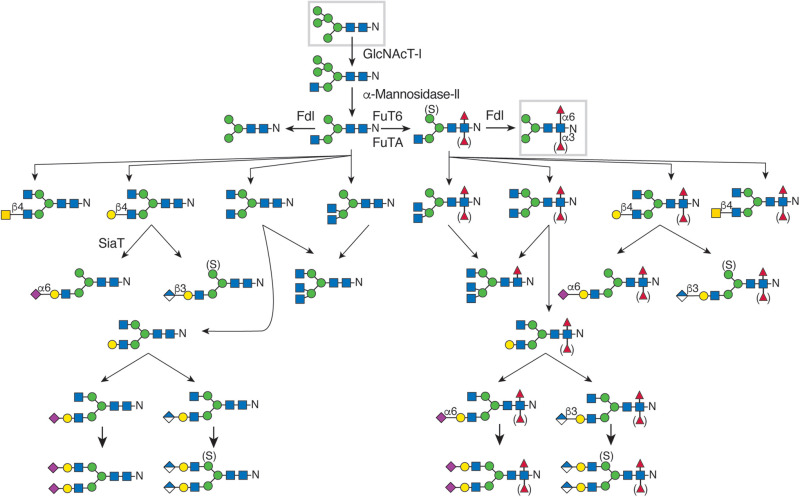
FIGURE 26.1.. N-Linked glycan diversity in Drosophila and other insects.

FIGURE 26.1.

N-Linked glycan diversity in Drosophila and other insects. N-Linked glycan processing after endoplasmic reticulum (ER) mannosidase trimming to the Man5GlcNAc2 structure is shown. (Gray boxes) The predominant N-linked glycans, Man5GlcNAc2 and Man3GlcNAc2Fuc, are found in all stages of Drosophila. N-linked glycan complexity is limited by the Fdl hexosaminidase and expanded by expression of uncharacterized branching N-acetylglucosaminyltransferase activities, undefined galactosyl-, N-acetylgalactosaminyl-, and glucuronosyl-transferase activities, and a single identified sialyltransferase activity. Core fucosylation occurs at C-6 and/or at C-3 of the reducing terminal N-acetylglucosamine residue, catalyzed by FucT6 or FucTA, respectively. Fucosylation at C-3 generates the neural-specific horseradish peroxidase (HRP) epitope. Sulfation of the α1-6-linked core mannose (Man) residue has been detected on all N-linked glycan classes except sialylated glycans in Drosophila and other insects.

Download Teaching Slide (PPTX, 11M)

From: Chapter 26, Arthropoda

Cover of Essentials of Glycobiology
Essentials of Glycobiology [Internet]. 4th edition.
Varki A, Cummings RD, Esko JD, et al., editors.
Cold Spring Harbor (NY): Cold Spring Harbor Laboratory Press; 2022.
Copyright © 2022 The Consortium of Glycobiology Editors, La Jolla, California; published by Cold Spring Harbor Laboratory Press; doi:10.1101/glycobiology.4e.26. All rights reserved.

The content of this book is licensed under a Creative Commons Attribution-NonCommercial-NoDerivs 4.0 Unported license. To view the terms and conditions of this license, visit https://creativecommons.org/licenses/by-nc-nd/4.0/

NCBI Bookshelf. A service of the National Library of Medicine, National Institutes of Health.