trehalose-6-phosphate synthase catalyzes the synthesis of alpha,alpha-1,1-trehalose-6-phosphate from glucose-6-phosphate using a uridine diphosphate-glucose donor
trehalose-6-phosphate synthase; Trehalose-6-Phosphate Synthase (TPS, EC 2.4.1.15) is a ...
288-827
2.06e-120
trehalose-6-phosphate synthase; Trehalose-6-Phosphate Synthase (TPS, EC 2.4.1.15) is a glycosyltransferase that catalyses the synthesis of alpha,alpha-1,1-trehalose-6-phosphate from glucose-6-phosphate using a UDP-glucose donor. It is a key enzyme in the trehalose synthesis pathway. Trehalose is a nonreducing disaccharide present in a wide variety of organisms and may serve as a source of energy and carbon. It is characterized most notably in insect, plant, and microbial cells. Its production is often associated with a variety of stress conditions, including desiccation, dehydration, heat, cold, and oxidation. This family represents the catalytic domain of the TPS. Some members of this domain family coexist with a C-terminal trehalose phosphatase domain.
:
Pssm-ID: 340820 [Multi-domain] Cd Length: 463 Bit Score: 382.32 E-value: 2.06e-120
Trehalose-6-phosphate phosphatase N-terminal helical bundle domain; This is the N-terminal ...
843-945
3.43e-34
Trehalose-6-phosphate phosphatase N-terminal helical bundle domain; This is the N-terminal domain found in trehalose-6-phosphate phosphatase (T6PP, EC 3.1.3.12) from parasitic nematodes such as Brugia malayi. In the model nematode Caenorhabditis elegans, T6PP is essential for survival due to the toxic effect(s) of the accumulation of trehalose 6-phosphate. T6PP has also been shown to be essential in Mycobacterium tuberculosis. The N-terminal domain composed of a three-helix bundle is similar in topology to the Microtubule Interacting and Transport (MIT) domains of the Vps4-like ATPases from Sulfolobus acidocaldarius. MIT domains are protein-interacting domains typically associated with multivesicular body formation, cytokinetic abscission, or viral budding. Mutational analysis indicate that deletion or mutation of the MIT-like domain is highly destabilizing to the enzyme.
:
Pssm-ID: 465806 Cd Length: 98 Bit Score: 126.55 E-value: 3.43e-34
trehalose-6-phosphate synthase; Trehalose-6-Phosphate Synthase (TPS, EC 2.4.1.15) is a ...
288-827
2.06e-120
trehalose-6-phosphate synthase; Trehalose-6-Phosphate Synthase (TPS, EC 2.4.1.15) is a glycosyltransferase that catalyses the synthesis of alpha,alpha-1,1-trehalose-6-phosphate from glucose-6-phosphate using a UDP-glucose donor. It is a key enzyme in the trehalose synthesis pathway. Trehalose is a nonreducing disaccharide present in a wide variety of organisms and may serve as a source of energy and carbon. It is characterized most notably in insect, plant, and microbial cells. Its production is often associated with a variety of stress conditions, including desiccation, dehydration, heat, cold, and oxidation. This family represents the catalytic domain of the TPS. Some members of this domain family coexist with a C-terminal trehalose phosphatase domain.
Pssm-ID: 340820 [Multi-domain] Cd Length: 463 Bit Score: 382.32 E-value: 2.06e-120
alpha,alpha-trehalose-phosphate synthase [UDP-forming]; This enzyme catalyzes the key, ...
288-764
1.27e-86
alpha,alpha-trehalose-phosphate synthase [UDP-forming]; This enzyme catalyzes the key, penultimate step in biosynthesis of trehalose, a compatible solute made as an osmoprotectant in some species in all three domains of life. The gene symbol OtsA stands for osmotically regulated trehalose synthesis A. Trehalose helps protect against both osmotic and thermal stresses, and is made from two glucose subunits. This model excludes glucosylglycerol-phosphate synthase, an enzyme of an analogous osmoprotectant system in many cyanobacterial strains. This model does not identify archaeal examples, as they are more divergent than glucosylglycerol-phosphate synthase. Sequences that score in the gray zone between the trusted and noise cutoffs include a number of yeast multidomain proteins in which the N-terminal domain may be functionally equivalent to this family. The gray zone also includes the OtsA of Cornyebacterium glutamicum (and related species), shown to be responsible for synthesis of only trace amounts of trehalose while the majority is synthesized by the TreYZ pathway; the significance of OtsA in this species is unclear (see Wolf, et al., ). [Cellular processes, Adaptations to atypical conditions]
Pssm-ID: 274112 Cd Length: 456 Bit Score: 289.56 E-value: 1.27e-86
Glycosyltransferase family 20; Members of this family belong to glycosyl transferase family 20. ...
287-764
3.46e-81
Glycosyltransferase family 20; Members of this family belong to glycosyl transferase family 20. OtsA (Trehalose-6-phosphate synthase) is homologous to regions in the subunits of yeast trehalose-6-phosphate synthase/phosphate complex,.
Pssm-ID: 425972 [Multi-domain] Cd Length: 471 Bit Score: 274.93 E-value: 3.46e-81
Trehalose-6-phosphate phosphatase N-terminal helical bundle domain; This is the N-terminal ...
843-945
3.43e-34
Trehalose-6-phosphate phosphatase N-terminal helical bundle domain; This is the N-terminal domain found in trehalose-6-phosphate phosphatase (T6PP, EC 3.1.3.12) from parasitic nematodes such as Brugia malayi. In the model nematode Caenorhabditis elegans, T6PP is essential for survival due to the toxic effect(s) of the accumulation of trehalose 6-phosphate. T6PP has also been shown to be essential in Mycobacterium tuberculosis. The N-terminal domain composed of a three-helix bundle is similar in topology to the Microtubule Interacting and Transport (MIT) domains of the Vps4-like ATPases from Sulfolobus acidocaldarius. MIT domains are protein-interacting domains typically associated with multivesicular body formation, cytokinetic abscission, or viral budding. Mutational analysis indicate that deletion or mutation of the MIT-like domain is highly destabilizing to the enzyme.
Pssm-ID: 465806 Cd Length: 98 Bit Score: 126.55 E-value: 3.43e-34
Cof subfamily of IIB subfamily of haloacid dehalogenase superfamily; This subfamily of ...
1165-1210
4.10e-04
Cof subfamily of IIB subfamily of haloacid dehalogenase superfamily; This subfamily of sequences falls within the Class-IIB subfamily (TIGR01484) of the Haloacid Dehalogenase superfamily of aspartate-nucleophile hydrolases. The use of the name "Cof" as an identifier here is arbitrary and refers to the E. coli Cof protein. This subfamily is notable for the large number of recent paralogs in many species. Listeria, for instance, has 12, Clostridium, Lactococcus and Streptococcus pneumoniae have 8 each, Enterococcus and Salmonella have 7 each, and Bacillus subtilus, Mycoplasma, Staphylococcus and E. coli have 6 each. This high degree of gene duplication is limited to the gamma proteobacteria and low-GC gram positive lineages. The profusion of genes in this subfamily is not coupled with a high degree of divergence, so it is impossible to determine an accurate phylogeny at the equivalog level. Considering the relationship of this subfamily to the other known members of the HAD-IIB subfamily (TIGR01484), sucrose and trehalose phosphatases and phosphomannomutase, it seems a reasonable hypothesis that these enzymes act on phosphorylated sugars. Possibly the diversification of genes in this subfamily represents the diverse sugars and polysaccharides that various bacteria find in their biological niches. The members of this subfamily are restricted almost exclusively to bacteria (one sequences from S. pombe scores above trusted, while another is between trusted and noise). It is notable that no archaea are found in this group, the closest relations to the archaea found here being two Deinococcus sequences. [Unknown function, Enzymes of unknown specificity]
Pssm-ID: 272905 [Multi-domain] Cd Length: 256 Bit Score: 43.80 E-value: 4.10e-04
Database: CDSEARCH/cdd Low complexity filter: no Composition Based Adjustment: yes E-value threshold: 0.01
References:
Wang J et al. (2023), "The conserved domain database in 2023", Nucleic Acids Res.51(D)384-8.
Lu S et al. (2020), "The conserved domain database in 2020", Nucleic Acids Res.48(D)265-8.
Marchler-Bauer A et al. (2017), "CDD/SPARCLE: functional classification of proteins via subfamily domain architectures.", Nucleic Acids Res.45(D)200-3.
of the residues that compose this conserved feature have been mapped to the query sequence.
Click on the triangle to view details about the feature, including a multiple sequence alignment
of your query sequence and the protein sequences used to curate the domain model,
where hash marks (#) above the aligned sequences show the location of the conserved feature residues.
The thumbnail image, if present, provides an approximate view of the feature's location in 3 dimensions.
Click on the triangle for interactive 3D structure viewing options.
Functional characterization of the conserved domain architecture found on the query.
Click here to see more details.
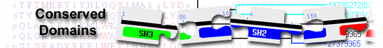
This image shows a graphical summary of conserved domains identified on the query sequence.
The Show Concise/Full Display button at the top of the page can be used to select the desired level of detail: only top scoring hits
(labeled illustration) or all hits
(labeled illustration).
Domains are color coded according to superfamilies
to which they have been assigned. Hits with scores that pass a domain-specific threshold
(specific hits) are drawn in bright colors.
Others (non-specific hits) and
superfamily placeholders are drawn in pastel colors.
if a domain or superfamily has been annotated with functional sites (conserved features),
they are mapped to the query sequence and indicated through sets of triangles
with the same color and shade of the domain or superfamily that provides the annotation. Mouse over the colored bars or triangles to see descriptions of the domains and features.
click on the bars or triangles to view your query sequence embedded in a multiple sequence alignment of the proteins used to develop the corresponding domain model.
The table lists conserved domains identified on the query sequence. Click on the plus sign (+) on the left to display full descriptions, alignments, and scores.
Click on the domain model's accession number to view the multiple sequence alignment of the proteins used to develop the corresponding domain model.
To view your query sequence embedded in that multiple sequence alignment, click on the colored bars in the Graphical Summary portion of the search results page,
or click on the triangles, if present, that represent functional sites (conserved features)
mapped to the query sequence.
Concise Display shows only the best scoring domain model, in each hit category listed below except non-specific hits, for each region on the query sequence.
(labeled illustration) Standard Display shows only the best scoring domain model from each source, in each hit category listed below for each region on the query sequence.
(labeled illustration) Full Display shows all domain models, in each hit category below, that meet or exceed the RPS-BLAST threshold for statistical significance.
(labeled illustration) Four types of hits can be shown, as available,
for each region on the query sequence:
specific hits meet or exceed a domain-specific e-value threshold
(illustrated example)
and represent a very high confidence that the query sequence belongs to the same protein family as the sequences use to create the domain model
non-specific hits
meet or exceed the RPS-BLAST threshold for statistical significance (default E-value cutoff of 0.01, or an E-value selected by user via the
advanced search options)
the domain superfamily to which the specific and non-specific hits belong
multi-domain models that were computationally detected and are likely to contain multiple single domains
Retrieve proteins that contain one or more of the domains present in the query sequence, using the Conserved Domain Architecture Retrieval Tool
(CDART).
Modify your query to search against a different database and/or use advanced search options